Review
. 2023 May-Jun;16(3):499-509.
doi: 10.5005/jp-journals-10005-2401.

Pediatric Maxillofacial Trauma: Insights into Diagnosis and Treatment of Mandibular Fractures in Pediatric Patients

Affiliations
Review

Pediatric Maxillofacial Trauma: Insights into Diagnosis and Treatment of Mandibular Fractures in Pediatric Patients

Elena Hofmann et al. Int J Clin Pediatr Dent. 2023 May-Jun.

Abstract

Aim: To assess the diagnostic and therapeutic approaches in pediatric mandibular fractures with regards to fracture pattern and localization.

Patients and methods: This retrospective study included patients under the age of 17 years that presented to the Department of Oral and Maxillofacial Surgery at Charité-Universitätsmedizin Berlin with mandibular fractures over an 11-year long period (2010-2020). Medical records were analyzed for age, gender, injury mechanism, clinical presentation, imaging modalities, fracture pattern, and fracture management. Statistical analyses included descriptive statistics, normality testing, and Mann-Whitney U tests.

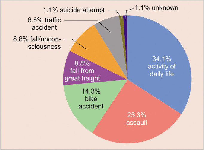
Results: A total of 91 pediatric patients (23 females and 68 males) presented with mandibular fractures. The majority of mandibular fractures occurred in patients aged 13-16 years (67.0%, n = 61). The main causes were activities of daily life (34.1%), followed by assault (25.3%). Malocclusion and pain upon mandibular joint compression were documented in 72.5% and 51.7% of patients, respectively. The most frequently applied radiological diagnostic tool was a panoramic X-ray (49.5%). The main fracture patterns were single (42.9%) and double fractures (48.4%). An age-adapted surgical approach using open reduction and internal fixation (ORIF) was the most frequent management (61.5%). A conservative approach was favored in cases of condylar head fractures. Resorbable plates were used in eight cases of ORIF (8.8%).

Conclusion: Treatment regimens should be carefully selected based on the unique anatomy of the pediatric patient with regards to centers of growth and dentition phase, to restore stomatognathic function and to maintain adequate skeletal growth and eruption of teeth.

Clinical significance: This study illustrates the challenges of mandibular fracture management in the pediatric patient.

How to cite this article: Hofmann E, Koerdt S, Heiland M, et al. Pediatric Maxillofacial Trauma: Insights into Diagnosis and Treatment of Mandibular Fractures in Pediatric Patients. Int J Clin Pediatr Dent 2023;16(3):499-509.

Keywords: Fracture management; Mandibular fracture; Pediatric maxillofacial trauma; Pediatric patient.


Conflict of interest statement

Source of support: Nil Conflict of interest: None

Figures

Figs 1A and B
Figs 1A and B
Pediatric mandibular fractures in relation to age and gender. (A) The numbers of female and male patients (n) are illustrated per age group. Age groups were based on the dentition phases. Patients in the age group 1–5 years were in the primary dentition phase, 6–12 years at the mixed dentition stage and 13–16 years at the permanent dentition phase. (B) The total number of patients with mandibular fractures per age (years) is displayed according to gender (n = 91)
Fig. 2
Fig. 2
Etiology of pediatric mandibular fractures. The proportion of injury mechanisms relative to the total number of patients 1–16 years of age with mandibular fractures is displayed (n = 91)
Figs 3A and B
Figs 3A and B
X-rays demonstrating exemplary mandibular fractures in pediatric patients. (A) Panoramic view displaying a parasymphysis fracture of the left mandible at the mixed dentition stage. (B) Postoperative panoramic X-ray following ORIF in a single parasymphysis mandibular fracture in a patient at the mixed dentition phase
Figs 4A to F
Figs 4A to F
Case presentation of a dislocated condylar neck fracture (Spiessl and Schroll, in an 8-year-old female patient. (A–C) Preoperative DVT scans demonstrating the angulated fracture of the right condylar neck (Spiessl and Schroll type IV).18 (D–F) Postoperative CT scans following ORIF using a preauricular extraoral surgical approach to the condylar region. Using short-acting muscle relaxants, a nerve stimulator is used to identify and preserve the facial nerve
Figs 5A and B
Figs 5A and B
Case presentation of a 5-year-old female patient with a single mandibular fracture of the left condylar head. (A) Panoramic view displaying a left condylar head fracture before orthodontic treatment using an activator appliance; (B) Postinterventional panoramic view 3 years after the trauma demonstrating sufficient reposition of the left condylar head
Figs 6A–H
Figs 6A–H
Case presentation of an 11-year-old male patient with a double mandibular fracture. Preoperative (A–D) CT scans and (E,F) Three-dimensional reconstruction images demonstrating the fractures localized at the left parasymphysis and the right condylar neck (Spiessl and Schroll type II). ORIF was performed to address the two fracture sites using resorbable osteosynthesis plates and screws. Postoperative panoramic view (G) 1-week and (H) 6 weeks after surgery
Figs 7A–E
Figs 7A–E
Case presentation of a 10-year-old male patient with a double mandibular fracture. Preoperative (A–C) CT scans display the fractures localized at the left parasymphysis site and the right condylar neck. ORIF was performed to address the parasymphysis fracture using resorbable osteosynthesis plates. Postoperative panoramic view (D) 1 day and (E) 5 months after surgery

References

    1. Rogan DT, Ahmed A. Pediatric facial fractures. Treasure Island (FL): StatPearls Publishing; 2021. [updated 2021 Jul 31]. Available at: - PubMed
    1. Boyette JR. Facial fractures in children. Otolaryngol Clin North Am. 2014;47(5):747–61. doi: 10.1016/j.otc.2014.06.008. - DOI - PubMed
    1. Sharma S, Vashistha A, Chugh A, et al. Pediatric mandibular fractures: a review. Int J Clin Pediatr Dent. 2009;2(2):1–5. doi: 10.5005/jp-journals-10005-1022. - DOI - PMC - PubMed
    1. Vyas RM, Dickinson BP, Wasson KL, et al. Pediatric facial fractures: current national incidence, distribution, and health care resource use. J Craniofac Surg. 2008;19(2):339–49; discussion 350. doi: 10.1097/SCS.0b013e31814fb5e3. - DOI - PubMed
    1. Thoren H, Iizuka T, Hallikainen D, et al. Different patterns of mandibular fractures in children. An analysis of 220 fractures in 157 patients. J Craniomaxillofac Surg. 1992;20(7):292–296. doi: 10.1016/s1010-5182(05)80398-1. - DOI - PubMed

LinkOut - more resources