

Enhanced 3D visualization of human fallopian tube morphology using a miniature optical coherence tomography catheter
- PMID: 37497483
- PMCID: PMC10368054
- DOI: 10.1364/BOE.489708
Enhanced 3D visualization of human fallopian tube morphology using a miniature optical coherence tomography catheter
Abstract
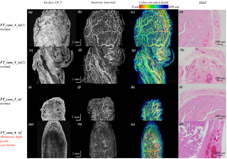
We demonstrate the use of our miniature optical coherence tomography catheter to acquire three-dimensional human fallopian tube images. Images of the fallopian tube's tissue morphology, vasculature, and tissue heterogeneity distribution are enhanced by adaptive thresholding, masking, and intensity inverting, making it easier to differentiate malignant tissue from normal tissue. The results show that normal fallopian tubes tend to have rich vasculature accompanied by a patterned tissue scattering background, features that do not appear in malignant cases. This finding suggests that miniature OCT catheters may have great potential for fast optical biopsy of the fallopian tube.
© 2023 Optica Publishing Group under the terms of the Optica Open Access Publishing Agreement.
Conflict of interest statement
The authors declare no conflicts of interest.
Figures


References
-
- Ducie J., Dao F., Considine M., Olvera N., Shaw P. A., Kurman R. J., Shih I. M., Soslow R. A., Cope L., Levine D. A., “Molecular analysis of high-grade serous ovarian carcinoma with and without associated serous tubal intra-epithelial carcinoma,” Nat. Commun. 8(1), 990 (2017).10.1038/s41467-017-01217-9 - DOI - PMC - PubMed
-
- Schmoeckel E., Odai-Afotey A. A., Schleißheimer M., Rottmann M., Flesken-Nikitin A., Ellenson L. H., Kirchner T., Mayr D., Nikitin A. Y., “LEF1 is preferentially expressed in the tubal-peritoneal junctions and is a reliable marker of tubal intraepithelial lesions,” Mod. Pathol. 30(9), 1241–1250 (2017).10.1038/modpathol.2017.53 - DOI - PMC - PubMed
-
- Madore W. J., De Montigny E., Deschênes A., Benboujja F., Leduc M., Mes-Masson A. M., Provencher D. M., Rahimi K., Boudoux C., Godbout N., “Morphologic three-dimensional scanning of fallopian tubes to assist ovarian cancer diagnosis,” J. Biomed. Opt 22(7), 076012 (2017).10.1117/1.JBO.22.7.076012 - DOI - PubMed
Grants and funding
LinkOut - more resources
Full Text Sources