. 2023 Jun 27;14(7):3775-3797.
doi: 10.1364/BOE.485090. eCollection 2023 Jul 1.

Fractal-based aberration-corrected full-field OCT

Affiliations

Fractal-based aberration-corrected full-field OCT

Yue Zhu et al. Biomed Opt Express. .

Abstract

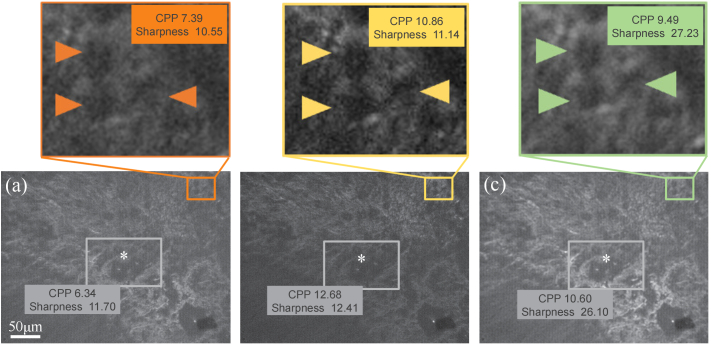
The Kolmogorov turbulence model has been validated as a quantitative 3D light scattering model of the inhomogeneous refraction index of biological tissue using full-field OCT (FF-OCT). A fractal-based computational compensation approach was proposed for correcting of depth-resolved aberrations with volumetric FF-OCT. First, the power-spectral density spectrum of the index inhomogeneities was measured by radial Fourier transformation of volumetric data. The spectrum's shape indicates the spatial correlation function and can be quantified as the fractal dimension of tissue. The defocusing correction matrix was built by applying fractal-based analysis as an image quality metric. For comparison, tissue-induced in-depth aberration models were built by phase compensation. After digital aberration correction of FF-OCT images, it enables extracting the temporal contrast indicating the sample dynamics in onion in mitosis and ex vivo mouse heart during delayed neuronal death. The proposed fractal-based contrast augmented images show subcellular resolution recording of dynamic scatters of the growing-up onion cell wall and some micro activities. In addition, low-frequency chamber and high-frequency cardiac muscle fibers from ex vivo mouse heart tissue. Therefore, the depth-resolved changes in fractal parameters may be regarded as a quantitative indicator of defocus aberration compensation. Also the enhanced temporal contrast in FF-OCT has the potential to be a label-free, non-invasive, and three-dimensional imaging tool to investigate sub-cellular activities in metabolism studies.


Conflict of interest statement

The authors declare no conflicts of interest.

Figures

Fig. 1.
Fig. 1.
Flowchart of Fourier-fractal refocusing algorithm.
Fig. 2.
Fig. 2.
Ray tracing model for defocus and spherical aberration.
Fig. 3.
Fig. 3.
En face histology-like tomographic images of (a) Esphagus; (b) Small intestine; (c) Spleen; (d) Kidney; (e) Liver; (f) Intestine; (g), (h) Heart at different depth; (i) Onion root tips undergoing mitosis.
Fig. 4.
Fig. 4.
(a) The Df compares linear with PSD curve fitting model in same tissue from Small intestine; (b) The Df compares in-focus with out-of-focus data in same tissue from small intestine; (c) The boxplot of refocused Df in seven mouse organs.
Fig. 5.
Fig. 5.
(a) In-depth fractal dimension curve comparison; (b),(c) FF-OCT images of fresh mouse liver tissue without and with applying refocusing, respectively.
Fig. 6.
Fig. 6.
(a), (b) Volume-based FF-OCT sectioned images of mouse heart without and with refocusing compensation.
Fig. 7.
Fig. 7.
En face tomographic images of fresh liver tissue (a) with amplitude reconstructions; (b) with tissue-induced aberration compensation; (c) with Fractal-based refocusing compensation
Fig. 8.
Fig. 8.
(a),(c) En face tomographic images of onion with the interval time of 1ms; (b),(d) Temporal contrast of corresponding FF-OCT images; (e) Colour bar of mean frequency for temporal contrast; (f),(h) En face tomographic images of the semi-dehydrated and the utterly dehydrated onion sample; (g),(i) Temporal contrast from the same ROI in the semi-dehydrated and utterly dehydrated onion sample.
Fig. 9.
Fig. 9.
Contrast-augmented FF-OCT images. (a) A series of FF-OCT images of fresh heart tissue; (b) Contrast augmented FF-OCT images of fresh heart tissue; (c) Colorbar of contrast augmented FF-OCT images.

References

    1. Beaurepaire E., Boccara A. C., Lebec M., Blanchot L., Saint-Jalmes H., “Full-field optical coherence microscopy,” Opt. Lett. 23(4), 244–246 (1998).10.1364/OL.23.000244 - DOI - PubMed
    1. Dubois A., Vabre L., Boccara A.-C., Beaurepaire E., “High-resolution full-field optical coherence tomography with a linnik microscope,” Appl. Opt. 41(4), 805–812 (2002).10.1364/AO.41.000805 - DOI - PubMed
    1. Scholler J., Groux K., Goureau O., Sahel J.-A., Fink M., Reichman S., Boccara C., Grieve K., “Dynamic full-field optical coherence tomography: 3d live-imaging of retinal organoids,” Light: Sci. Appl. 9(1), 140 (2020).10.1038/s41377-020-00375-8 - DOI - PMC - PubMed
    1. Tomczewski S., Wegrzyn P., Borycki D., Auksorius E., Wojtkowski M., Curatolo A., “Light-adapted flicker optoretinograms captured with a spatio-temporal optical coherence-tomography (stoc-t) system,” Biomed. Opt. Express 13(4), 2186–2201 (2022).10.1364/BOE.444567 - DOI - PMC - PubMed
    1. Auksorius E., Borycki D., Wegrzyn P., Sikorski B. L., Lizewski K., Zickiene I., Rapolu M., Adomavicius K., Tomczewski S., Wojtkowski M., “Spatio-temporal optical coherence tomography provides full thickness imaging of the chorioretinal complex,” iScience 25(12), 105513 (2022).10.1016/j.isci.2022.105513 - DOI - PMC - PubMed

LinkOut - more resources