

Relevance and utility of the in-vivo and ex-vivo optical properties of the skin reported in the literature: a review [Invited]
- PMID: 37497524
- PMCID: PMC10368038
- DOI: 10.1364/BOE.493588
Relevance and utility of the in-vivo and ex-vivo optical properties of the skin reported in the literature: a review [Invited]
Abstract
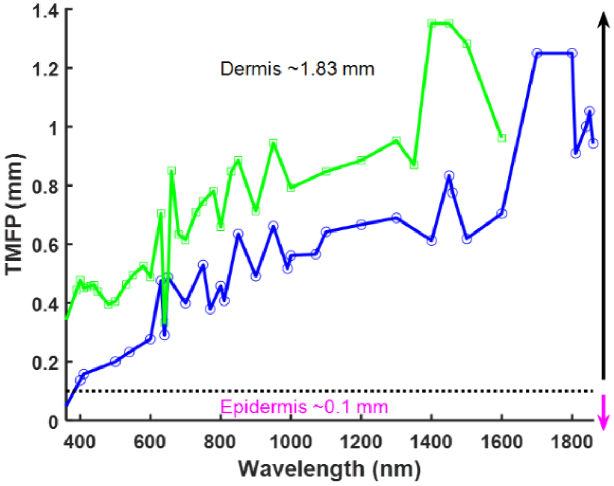
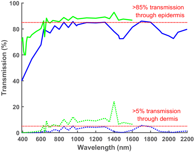
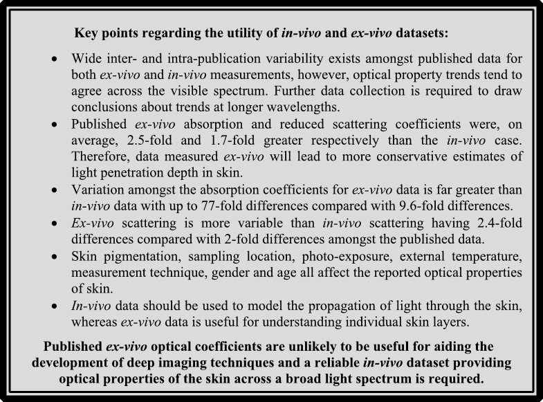
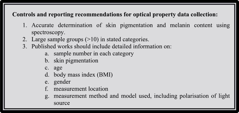
Imaging non-invasively into the human body is currently limited by cost (MRI and CT scan), image resolution (ultrasound), exposure to ionising radiation (CT scan and X-ray), and the requirement for exogenous contrast agents (CT scan and PET scan). Optical imaging has the potential to overcome all these issues but is currently limited by imaging depth due to the scattering and absorption properties of human tissue. Skin is the first barrier encountered by light when imaging non-invasively, and therefore a clear understanding of the way that light interacts with skin is required for progress on optical medical imaging to be made. Here we present a thorough review of the optical properties of human skin measured in-vivo and compare these to the previously collated ex-vivo measurements. Both in-vivo and ex-vivo published data show high inter- and intra-publication variability making definitive answers regarding optical properties at given wavelengths challenging. Overall, variability is highest for ex-vivo absorption measurements with differences of up to 77-fold compared with 9.6-fold for the in-vivo absorption case. The impact of this variation on optical penetration depth and transport mean free path is presented and potential causes of these inconsistencies are discussed. We propose a set of experimental controls and reporting requirements for future measurements. We conclude that a robust in-vivo dataset, measured across a broad spectrum of wavelengths, is required for the development of future technologies that significantly increase the depth of optical imaging.
Published by Optica Publishing Group under the terms of the Creative Commons Attribution 4.0 License. Further distribution of this work must maintain attribution to the author(s) and the published article’s title, journal citation, and DOI.
Conflict of interest statement
The authors declare no conflicts of interest.
Figures
Similar articles
-
Effect of skin color on optical properties and the implications for medical optical technologies: a review.J Biomed Opt. 2024 Jan;29(1):010901. doi: 10.1117/1.JBO.29.1.010901. Epub 2024 Jan 24. J Biomed Opt. 2024. PMID: 38269083 Free PMC article. Review.
-
Optical clearing of skin using flash lamp-induced enhancement of epidermal permeability.Lasers Surg Med. 2006 Oct;38(9):824-36. doi: 10.1002/lsm.20392. Lasers Surg Med. 2006. PMID: 17044094
-
Spectra from 2.5-15 microm of tissue phantom materials, optical clearing agents and ex vivo human skin: implications for depth profiling of human skin.Phys Med Biol. 2003 Jan 21;48(2):N15-24. doi: 10.1088/0031-9155/48/2/402. Phys Med Biol. 2003. PMID: 12587910
-
Early assessment of burn severity in human tissue ex vivo with multi-wavelength spatial frequency domain imaging.Toxicol In Vitro. 2018 Oct;52:251-254. doi: 10.1016/j.tiv.2018.05.015. Epub 2018 Jun 1. Toxicol In Vitro. 2018. PMID: 29859991 Free PMC article.
-
Optical coherence tomography in dermatology.G Ital Dermatol Venereol. 2015 Oct;150(5):603-15. Epub 2015 Jul 1. G Ital Dermatol Venereol. 2015. PMID: 26129683 Review.
Cited by
-
Real Index of Refraction of Normal and Cancerous Axillary Lymph Nodes in Breast Cancer Patients: Results from an Experimental Study.J Pers Med. 2025 Feb 18;15(2):71. doi: 10.3390/jpm15020071. J Pers Med. 2025. PMID: 39997348 Free PMC article.
-
Enhancing tumor's skin photothermal therapy using Gold nanoparticles : a Monte Carlo simulation.Lasers Med Sci. 2024 May 16;39(1):130. doi: 10.1007/s10103-024-04072-5. Lasers Med Sci. 2024. PMID: 38750285
-
Depth-dependent fluence compensation without a priori knowledge of tissue composition for quantitative ultrasound-guided photoacoustic imaging.J Biomed Opt. 2025 Jul;30(7):076005. doi: 10.1117/1.JBO.30.7.076005. Epub 2025 Jul 12. J Biomed Opt. 2025. PMID: 40655940 Free PMC article.
-
Effect of skin color on optical properties and the implications for medical optical technologies: a review.J Biomed Opt. 2024 Jan;29(1):010901. doi: 10.1117/1.JBO.29.1.010901. Epub 2024 Jan 24. J Biomed Opt. 2024. PMID: 38269083 Free PMC article. Review.
-
Age dependency of the reduced scattering coefficient and viscoelastic character, and their relationship in Japanese female skin.Biomed Opt Express. 2024 Jul 19;15(8):4775-4785. doi: 10.1364/BOE.527858. eCollection 2024 Aug 1. Biomed Opt Express. 2024. PMID: 39346985 Free PMC article.
References
-
- Bashkatov A. N., Berezin K. V., Dvoretskiy K. N., Chernavina M. L., Genina E. A., Genin V. D., Kochubey V. I., Lazareva E. N., Pravdin A. B., Shvachkina M. E., Timoshina P. A., Tuchina D. K., Yakovlev D. D., Yakovlev D. A., Yanina I. Y., Zhernovaya O. S., Tuchin V. V., “Measurement of tissue optical properties in the context of tissue optical clearing,” J. Biomed. Opt. 23(09), 091416 (2018).10.1117/1.JBO.23.9.091416 - DOI - PubMed
-
- “Anatomy of the Skin,” https://www.utmb.edu/pedi_ed/CoreV2/Dermatology/page_03.htm.
Publication types
LinkOut - more resources
Full Text Sources