

Patellar malalignment correlates with increased pain and increased synovial stress hormone levels-A cross-sectional study
- PMID: 37498905
- PMCID: PMC10374142
- DOI: 10.1371/journal.pone.0289298
Patellar malalignment correlates with increased pain and increased synovial stress hormone levels-A cross-sectional study
Abstract
Purpose: Risk factors for the development of pain in the context of knee osteoarthritis (KOA) remain unclear. Radiological findings often do not correlate with clinical findings, so other pathomechanisms in the development and perception of pain must play a role. The purpose of this study is to investigate the correlation of increased sympathetic nervous system (SNS) activity (measured by subjective and objective chronic stress parameters) with KOA severity, patellofemoral malalignment, and pain.
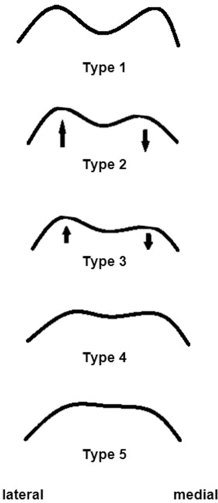
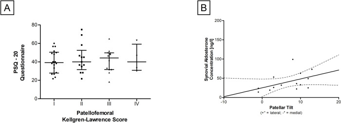
Methods: 47 patients with KOA were assessed. Radiological measurements of tibiofemoral and patellofemoral parameters (Kellgren-Lawrence-score, patellar tilt (PT), Caton-Deschamps-Index and Hepp´s classification) were performed and correlated with knee-specific questionnaires (WOMAC®, KSS©) and chronic stress questionnaires (PSQ-20). Additionally, parameters associated with chronic stress were quantified in synovial fluid and serum samples from patients.
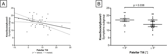
Results: PT correlated significantly with Caton-Deschamps-Index (r = 0.394,p = 0.006) and with medial patellofemoral joint space (r = 0.516,p<0.001). In addition, asymmetric trochlear groove (Hepp's classification > II) was associated with significantly higher PT values (p = 0.014). A negative correlation between PT and KSS©-symptoms subgroup was found (r = -0.340,p = 0.024). Patients with PT<5° had significantly higher scores in the Knee Society Score©-symptoms subgroup (p = 0.038). A positive and significant correlation between synovial aldosterone levels and PT was observed (r = 0.548,p = 0.042).
Conclusion: The results of this study indicate that patellar malalignment might correlate with increased pain. The previous specification of standard PT values must be reconsidered as even low PT values seem to play a role in the occurrence of patellofemoral osteoarthritis symptoms. Lower PT values might lead to aggravated symptoms in patients with KOA due to a narrow medial patellofemoral joint space. In addition, PT might induce the release of synovial stress biomarkers and thus contribute to the progression of KOA.
Copyright: © 2023 Brenneis et al. This is an open access article distributed under the terms of the Creative Commons Attribution License, which permits unrestricted use, distribution, and reproduction in any medium, provided the original author and source are credited.
Conflict of interest statement
The authors have declared that no competing interests exist.
Figures


Similar articles
-
Correlation between severity of knee joint osteoarthritis and alignment of patellofemoral and patellar height on radiographs.Chin Med J (Engl). 2025 Apr 20;138(8):947-952. doi: 10.1097/CM9.0000000000003392. Epub 2024 Nov 15. Chin Med J (Engl). 2025. PMID: 39602320 Free PMC article.
-
Correlation between varus knee malalignment and patellofemoral osteoarthritis.Knee Surg Sports Traumatol Arthrosc. 2016 Jan;24(1):176-81. doi: 10.1007/s00167-014-3360-3. Epub 2014 Oct 2. Knee Surg Sports Traumatol Arthrosc. 2016. PMID: 25274097
-
Correction of Patellofemoral Malalignment With Patellofemoral Arthroplasty.J Arthroplasty. 2017 Dec;32(12):3598-3602. doi: 10.1016/j.arth.2017.06.048. Epub 2017 Jul 8. J Arthroplasty. 2017. PMID: 28735802
-
[Correlation analysis between imaging classification of varus knee osteoarthritis and axis angle of tibiofemoral and patellofemoral joints].Zhongguo Gu Shang. 2023 Apr 25;36(4):364-70. doi: 10.12200/j.issn.1003-0034.2023.04.013. Zhongguo Gu Shang. 2023. PMID: 37087627 Chinese.
-
Osteoarthritic knees have a highly variable patellofemoral alignment: a systematic review.Knee Surg Sports Traumatol Arthrosc. 2021 Feb;29(2):483-490. doi: 10.1007/s00167-020-05928-3. Epub 2020 Mar 12. Knee Surg Sports Traumatol Arthrosc. 2021. PMID: 32162047
Cited by
-
Comparison of Inflammatory Biomarkers in Females with and Without Patellofemoral Pain and Associations with Patella Position, Hip and Knee Kinematics, and Pain.Biomedicines. 2025 Mar 20;13(3):761. doi: 10.3390/biomedicines13030761. Biomedicines. 2025. PMID: 40149737 Free PMC article.
-
Complement 3 and 4 impact in osteoarthritis.Biomark Med. 2025 Feb;19(3):81-90. doi: 10.1080/17520363.2024.2409062. Epub 2025 Feb 2. Biomark Med. 2025. PMID: 39893562
References
MeSH terms
Substances
LinkOut - more resources
Full Text Sources
