. 2023 Jul 28;381(6656):436-443.
doi: 10.1126/science.ade6967. Epub 2023 Jul 27.

In vivo hematopoietic stem cell modification by mRNA delivery

Affiliations

In vivo hematopoietic stem cell modification by mRNA delivery

Laura Breda et al. Science. .

Erratum in

Abstract

Hematopoietic stem cells (HSCs) are the source of all blood cells over an individual's lifetime. Diseased HSCs can be replaced with gene-engineered or healthy HSCs through HSC transplantation (HSCT). However, current protocols carry major side effects and have limited access. We developed CD117/LNP-messenger RNA (mRNA), a lipid nanoparticle (LNP) that encapsulates mRNA and is targeted to the stem cell factor receptor (CD117) on HSCs. Delivery of the anti-human CD117/LNP-based editing system yielded near-complete correction of hematopoietic sickle cells. Furthermore, in vivo delivery of pro-apoptotic PUMA (p53 up-regulated modulator of apoptosis) mRNA with CD117/LNP affected HSC function and permitted nongenotoxic conditioning for HSCT. The ability to target HSCs in vivo offers a nongenotoxic conditioning regimen for HSCT, and this platform could be the basis of in vivo genome editing to cure genetic disorders, which would abrogate the need for HSCT.


Conflict of interest statement

S.R. is a member of scientific advisory board of Ionis Pharmaceuticals, Meira GTx, Vifor and Disc Medicine. H.P., and D.W. are scientific founders and hold equity in Capstan Therapeutics. Y.K.T. and B.L.M. are employees and hold equity in Acuitas Therapeutics. D.W. and H.P. receive research support from BioNTech. L.B., T.E.P., M.P.T., S.R., and H.P. are inventors (University of Pennsylvania) on a patent for the in vivo gene editing of hematopoietic stem cells using targeted LNP for hemoglobinopathies (US Provisional Patent Application 63/499,580 filed 2 May 2023). L.B., T.E.P., M.P.T., S.R., and H.P. are inventors (University of Pennsylvania) on a patent for HSC-targeted LNP-mRNA for conditioning before HSC transplant (US Provisional Patent Application 046483-6260 filed 9 December 2022). D.W., and H.P. are inventors (University of Pennsylvania) on a patent on the compositions and methods for targeting lipid nanoparticle mRNA therapeutics to stem cells (US Provisional Patent Application 63/182,639 filed 30 April 2021, WIPO Patent Application PCT/US2022/026933). In accordance with the University of Pennsylvania policies and procedures and our ethical obligations as researchers, D.W. and H.P. are named on additional patents that describe the use of nucleoside-modified mRNA and targeted LNPs as platforms to deliver therapeutic proteins and vaccines. These interests have been fully disclosed to the University of Pennsylvania, and approved plans are in place for managing any potential conflicts arising from licensing these patents.

Figures

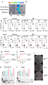
Fig. 1
Fig. 1. In vitro targeting of whole bone marrow or hematopoietic progenitors (lin-) cells incubated with LNPs encapsulating luciferase (CD117/LNP-Luc) or Cre recombinase (CD117/LNP-Cre) mRNAs.
(A) Luciferase activity normalized by total protein in whole bone marrow cells incubated with varying doses (indicated on x-axis) of targeted or control LNP-Luc for 18 hours in vitro. Data indicate mean +/− SD of n=3 replicate experiments. P values are from Dunnett’s multiple comparison after two-way ANOVA. **** p<0.0001. (B) LNP-Luc treatment of Lineage negative (Lin-) bone marrow cells (N=3). Data indicate mean +/− SD of n=3 replicate experiments. P values are from Dunnett’s multiple comparison after two-way ANOVA. **** p<0.0001. (C-G) Assessment of ZsGreen+ reporter induction after CD117/LNP-Cre treatment in Ai6 bone marrow (BM) cells triggered by removal of loxP flanked stop cassette by Cre. Treatment of (C, E, G) bone marrow cells or (D, F) Lin-BM cells at doses and culture intervals stated in figure. (D, F) Lin-Sca1+cKit+ (LSK) subset shown when treating Lin-cells. No difference between CD117/LNP-Cre editing in Lin- cells treated with 0.1 and 0.5 or 0.5 and 1μg;. In C-G, data represent mean +/− SD of n=3 replicate experiments. P values are from Dunnett’s multiple comparison after two-way ANOVA. Specifically, in C-G, ** p<0.01, *** p<0.001 **** p<0.0001.
Fig. 2
Fig. 2. CD117/LNP-Cre treatment ex vivo leads to near complete tdTomato gene editing upon transplantation
(A) Percent tdTomato marking in myeloid (Gr1+), (B) lymphoid (CD3+, left and B220+, right) cells measured at 16 weeks post HSCT in lethally irradiated congenic CD45.1 recipients receiving Ai14 bone marrow treated ex vivo with 0.1 μg (and 1 μg of control IgG/LNP-Cre or CD117/LNP-Cre. In A and B data represent mean +/− SEM of n=4 (for IgG/LNP-cre at 1μg only) or n=5 experimental animals per cohort. P-values are from Tukey’s multiple comparison test after one-way ANOVA. In A**** p<0.0001. In B *p<0.05, **** p<0.0001. (C) Kinetic analysis of erythroid editing measured up to 16 weeks post HSCT. Data represent mean +/− SD of n=4 or 5 experimental animals per cohort. (D) tdTomato marking in in the bone marrow (BM) and BM subsets: c-Kit+ (Lin-c-Kit+), LSK (Lin-c-Kit+Sca1+), and LT-HSC (LSK CD150+CD48-). Data represent mean +/− SEM of n=4 or 5 experimental animals per cohort (same animals as in A and B). P-values are Tukey’s multiple comparison test after one-way ANOVA. *** p<0.001, and **** p<0.0001. (E) Colony forming unit assay from Ai14 bone marrow treated ex vivo with 0.1 μg or 1 μg of control IgG/LNP-Cre or CD117/LNP-Cre formulations or untreated. Semi-quantitative PCR of (F) bone marrow and (G) spleen genomic DNA isolates from the groups in A-C at 4 months post BMT. **271bp Cre-recombinase edited gDNA region, *1142bp unedited region are indicated.
Fig. 3
Fig. 3. CD117/LNP-Cre formulations lead to over 50% tdTomato marking in LT-HSC after in vivo injection
(A) Biodistribution of i.v. injection of 1 μg of targeted LNP-mRNA expression in vivo by luminescence imaging at 24 hours. A representative sample set of dissected mouse organs were analyzed 5 min after the administration of D-luciferin. tdTomato+ cell frequency in peripheral blood (B) myeloid (Gr1+) and (C) lymphoid cells (CD3+ [T-cells], B220+ [B-cells]) and in (D) bone marrow (BM) subsets (c-Kit, Lin-c-Kit+ subset, LSK, Lin-c-Kit+Sca1+, SLAM/LT-HSC, LSK CD150+ CD48-) at 4 months after 5 μg of CD117/LNP-Cre or control IgG/LNP-Cre. In B, C, and D data represent mean +/− SEM of n=5 experimental animals per cohort. P-values are reported from paired t-test. ** p<0.01, *** p<0.001, and **** p<0.0001. tdTomato+ cell frequency in peripheral blood (E) myeloid and (F) lymphoid cells, and in (G) bone marrow subsets at 4 months after 5 or 1 μg of CD117/LNP-Cre. In E, F, and G data represent mean +/− SEM of n=7 (1μg) and 5 (5μg) experimental animals per cohort. P-values are reported from t-test. *** p<0.001, and **** p<0.0001. Edited RBC frequency over time in Ai9 mice treated in vivo with (H) 5 μg of CD117/LNP-Cre or control IgG/LNP-Cre or with (I) 1 or 5 μg of CD117/LNP-Cre. In H data represent mean +/− SD of n=5 experimental animal per cohort. P-values are reported from paired t-test. **** p<0.0001. In I data represent mean +/− SD of n=7 (1μg) and 5 (5μg) experimental animals per cohort. P-values are reported from t-test. **** p<0.0001. J) Colony forming unit assay from bone marrow at 4 months after in vivo treatment with 5 μg control-IgG/LNP-Cre (top), no treatment (middle), or 5 μg CD117/LNP-Cre (bottom). Semi-quantitative PCR of (K) bone marrow and (L) spleen genomic DNA isolates from the groups in A-C at 4 months post BMT. **271bp Cre-recombinase edited gDNA region, *1142bp unedited region are indicated.
Fig. 4
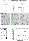
Fig. 4. Base editing of the E6V sickle cell mutation with Human CD117 targeted LNP
(A) Representative reverse-phase (RP) HPLC chromatograms of in vitro differentiated sickle cell disease erythroid progenitor lysates after treatment with anti-human CD117 (hCD117)/LNPNRCH cas9 ABE-8e mRNA and hCD117/LNP gRNA. Base editing yields non-pathogenic HbbG-MakassarG), which elutes before HbbS (pathogenic, βS) and the α-globin protein (α). % shown is βG/(βGs) *100. (B) Representative images of sickling of in vitro differentiated erythroid progenitors under hypoxic conditions at the treatments in (A). Arrowheads indicate sickled morphology. Scale bar 20 microns. (C) % sickled cells from unedited and edited (varying mRNA doses) sickling assays. Data represent mean +/− SD of n=10 high powered fields (hpf) (unedited specimens) and n=30 hpf (edited specimens). P-values are reported from unpaired t-test. **** p<0.0001. (D) Correlation of %βG by RP-HPLC (protein) to base edited allele frequency (DNA).
Fig. 5
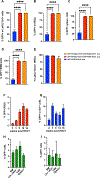
Fig. 5. HSC depletion and transplantation conditioning with CD117/LNP-PUMA
(A) GFP+ granulocytes and (B) RBC in peripheral blood, as well as %GFP+ (C) CD45+ splenocytes and (D) BM cells of C57BL/6 CD45.1 chimeras competitively transplanted with indicated proportion of GFP+ C57BL/6 BM untreated and C57/BL6 (GFP-) BM treated with CD117/LNP-PUMA. Data represent mean +/− SD of n=4 (recipients of a 25:75 ratio of GFP:C57/BL6+CD117/LNP-PUMA BM), n=8 (recipients of a 50:50 ratio of GFP:C57/BL6+CD117/LNP-PUMA BM), and n=4 (recipients of a 50:50 ratio of GFP:C57/BL6 untreated BM) experimental animals per cohort. P-values calculated by Dunnett’s multiple comparison test after one-way ANOVA.**** p<0.0001. (E) Donor chimerism 4 months post HSCT. Chimerism calculated as CD45.2%/(CD45.1%+CD45.2%). Data represent mean +/− SD of the same cohorts indicated in A-C. One-way ANOVA not significant (p>0.05). (F) Granulocyte, (G) RBC, and (H) hematopoietic cells of the BM, BM subsets, and spleen in recipients conditioned with 0.05 mg/kg CD117/LNP-PUMA and receiving 10×106 GFP+ C57BL/6 BM cells at 6.5 days post treatment. Data in F-H represent mean +/− SD of n=3 recipient animals. Levels of GFP+ granulocytes and RBC in unconditioned controls (N=2) were nearly undetected (0.06±0.03 and 0.05±0.02, respectively) 2 months after BMT. (I) Persistence upon secondary transplantation of CD117/LNP-PUMA conditioned GFP+ donor BM in lethally irradiated congenic mice. Data represent mean +/− SD of n=8 recipient animals generated from 3 primary chimeras.

Comment in

References

    1. Kent D et al. , Regulation of hematopoietic stem cells by the steel factor/KIT signaling pathway. Clin Cancer Res 14, 1926–1930 (2008). - PubMed
    1. Yee NS, Hsiau CW, Serve H, Vosseller K, Besmer P, Mechanism of down-regulation of c-kit receptor. Roles of receptor tyrosine kinase, phosphatidylinositol 3’-kinase, and protein kinase C. J Biol Chem 269, 31991–31998 (1994). - PubMed
    1. Weissman D, Kariko K, mRNA: Fulfilling the Promise of Gene Therapy. Mol Ther 23, 1416–1417 (2015). - PMC - PubMed
    1. Kariko K, Buckstein M, Ni H, Weissman D, Suppression of RNA recognition by Toll-like receptors: the impact of nucleoside modification and the evolutionary origin of RNA. Immunity 23, 165–175 (2005). - PubMed
    1. Kariko K, Muramatsu H, Ludwig J, Weissman D, Generating the optimal mRNA for therapy: HPLC purification eliminates immune activation and improves translation of nucleoside-modified, protein-encoding mRNA. Nucleic Acids Res 39, e142 (2011). - PMC - PubMed