. 2023 Oct 15:1817:148496.
doi: 10.1016/j.brainres.2023.148496. Epub 2023 Jul 25.

Tight control of the APP-Mint1 interaction in regulating amyloid production

Affiliations

Tight control of the APP-Mint1 interaction in regulating amyloid production

Shawna M Henry et al. Brain Res. .

Abstract

Generation of amyloid-β (Aβ) peptides through the proteolytic processing of the amyloid precursor protein (APP) is a pathogenic event in Alzheimer's disease (AD). APP is a transmembrane protein and endocytosis of APP mediated by the YENPTY motif is a key step in Aβ generation. Mints, a family of cytosolic adaptor proteins, directly bind to the YENPTY motif of APP and facilitate APP trafficking and processing. Here, we generated and examined two Mint1 mutants, Tyr633Ala of Mint1 (Mint1Y633A) that enhanced APP binding, and Tyr549Ala and Phe610Ala mutant (Mint1Y549A/F610A), that reduced APP binding. We investigated how perturbing the APP-Mint1 interaction through these Mint1 mutants alter APP and Mint1 cellular dynamics and Mint1's interaction with its other binding partners. We found that Mint1Y633A increased binding affinity specifically for APP and presenilin1 (catalytic subunit of γ-secretase), that subsequently enhanced APP endocytosis in primary murine neurons. Conversely, Mint1Y549A/F610A exhibited reduced APP affinity and Aβ secretion. The effect of Mint1Y549A/F610A on Aβ release was greater compared to knocking down all three Mint proteins supporting the APP-Mint1 interaction is a critical factor in Aβ production. Altogether, this study highlights the potential of targeting the APP-Mint1 interaction as a therapeutic strategy for AD.

Keywords: APP; Alzheimer’s disease; Amyloid; Mint1.


Conflict of interest statement

Declaration of Competing Interest The authors declare that they have no known competing financial interests or personal relationships that could have appeared to influence the work reported in this paper.

Figures

Fig. 1.
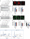
Fig. 1.. Biochemical analysis of Mint1 mutants with APP family of proteins and interacting partners.
(A-C) HEK293T cells were transfected GFP-Mint1WT, GFP-Mint1Y633A, or GFP-Mint1Y549A/F610A alone or co-transfected with APP, APLP1 or APLP2. Cell lysates were immunoprecipitated (IP) with GFP antibody and immunoblotted for APP, APLP1, APLP2, GFP, and tubulin. The amount of immunoprecipitated was normalized to the amount of precipitated Mint1 and shown as percent Mint1WT control. Data are expressed as the mean ± SEM (n = 4 independent experiments). Statistical significance was evaluated using one-way ANOVA with Sidak’s multiple comparison test, ***p = 0.00012 and **** p = 0.00009. (D-G) Fluorescence polarization (FP) saturation curves of the binding of APP peptide (D), PS1 (E), Nrxn1 (F), VGCC2.2 to recombinantly expressed Mint1WT, Mint1Y633A, or Mint1Y549A/F610A mutations. (H-K) Affinity fold-change to APP peptide (H), PS1 (I), Nrxn1 (J), VGCC2.2 (K) toward Mint1WT, Mint1Y633A, or Mint1Y549A/F610A mutations obtained in a FP assay. (L) Summary comparison of fold change for APP and Mint1 interacting partners toward Mint1WT and mutants (n = 1 independent experiment with 3 biological replicates). Statistical significance was evaluated using one-way ANOVA with Sidak’s multiple comparison test, *p < 0.05 and ***p < 0.0001.
Fig. 2.
Fig. 2.. Cellular localization of Mint1 mutants in primary neurons.
(A) Primary murine neurons that lacked endogenous Mint1 and infected with GFP-Mint1WT, GFP-Mint1Y633A, or GFP-Mint1Y549A/F610A were immunolabeled with GFP and cis-Golgi marker GM130. Representative images show GFP-Mint1 (green) and Golgi staining (red). Scale bar = 10 μm. (B) Co-localization of GFP-Mint1 with GM130 was quantified. (C) Percentage of neurons exhibiting a ribbon or condensed Golgi phenotype. Same neurons as analyzed in panel B. (D) Representative images show GFP-Mint1 (green) and APP staining (red). Scale bar for soma = 10 μm and processes = 5 μm. (E-F) Co-localization of GFP-Mint1 with APP was quantified in soma and processes. (G) Representative images show GFP-Mint1 (green) and synapsin staining (red). Scale bar for soma = 10 μm and processes = 5 μm. (H-I) Co-localization of GFP-Mint1 with synapsin was quantified in soma and processes. Data are expressed as the mean ± SEM (n = 1 independent experiment, number at the bottom of each bar represents number of neurons analyzed from 3–4 coverslips per condition). Statistical significance was evaluated using one-way ANOVA with Sidak’s multiple comparison test, *p ≤ 0.05, **p ≤ 0.01, ***p ≤ 0.001.
Fig. 3.
Fig. 3.. Mint1 mutants alter APP endocytosis and Aβ production in primary neurons.
Mint triple-floxed neurons were infected with inactive lentiviral Cre recombinase (ΔCre) or active Cre recombinase to knockdown Mints 1–3 and rescued with GFP-Mint1WT, GFP-Mint1Y633A, or GFP-Mint1Y549A/F610A lentivirus. (A) Representative immunoblot analysis of neuronal lysates immunoblotted for GFP, individual Mint proteins, APP and tubulin serves as a loading control. (B) Representative immunoblots of neuronal lysates immunoblotted for APP, APP-CTF and GAPDH as a loading control. (C) Representative immunoblots of neuronal lysates immunoblotted for sAPPα, sAPPα and GAPDH. (D) Representative images showing internalized APP (red) in both the soma (top) and processes (bottom). Scale bars: soma = 10 μm; process = 5 μm. (E-F) Quantification of the amount of internalized APP using corrected total cell fluorescence in the neuronal soma and processes, expressed as percent Mint1WT. (G) Representative images showing cell surface APP (green) in both the soma (top) and processes (bottom). (H-I) Quantification of the amount of cell surface APP using corrected total cell fluorescence in the neuronal soma and processes, expressed as percent Mint1WT. Data are expressed as the mean ± SEM (n = 1–2 independent experiment, number at the bottom of each bar represents number of neurons analyzed). (J) Aβ42 ELISA quantification of conditioned media from neurons cultured from Mint triple-floxed carrying the human APPswePS1ΔE9 transgene. Data were normalized to ΔCre control, and expressed as the mean ± SEM (n = 1 independent experiment with 3 biological replicates). Statistical significance was evaluated using one-way ANOVA with Sidak’s multiple comparison test, *p ≤ 0.05, **p ≤ 0.01, ***p ≤ 0.001.

References

    1. Bartling CRO, Jensen TMT, Henry SM, Colliander AL, Sereikaite V, Wenzler M, Jain P, Maric HM, Harpsoe K, Pedersen SW, Clemmensen LS, Haugaard-Kedstrom LM, Gloriam DE, Ho A, Stromgaard K, 2021. Targeting the APP-Mint2 Protein-Protein Interaction with a Peptide-Based Inhibitor Reduces Amyloid-beta Formation. J Am Chem Soc. 143, 891–901. - PMC - PubMed
    1. Biederer T, Cao X, Sudhof TC, Liu X, 2002a. Regulation of APP-dependent transcription complexes by Mint/X11s: differential functions of Mint isoforms. J Neurosci. 22, 7340–51. - PMC - PubMed
    1. Caster AH, Kahn RA, 2013. Recruitment of the Mint3 adaptor is necessary for export of the amyloid precursor protein (APP) from the Golgi complex. J Biol Chem. 288, 28567–80. - PMC - PubMed
    1. Chaufty J, Sullivan SE, Ho A, 2012. Intracellular amyloid precursor protein sorting and amyloid-beta secretion are regulated by Src-mediated phosphorylation of Mint2. J Neurosci. 32, 9613–25. - PMC - PubMed
    1. Cirrito JR, Kang JE, Lee J, Stewart FR, Verges DK, Silverio LM, Bu G, Mennerick S, Holtzman DM, 2008. Endocytosis is required for synaptic activity-dependent release of amyloid-beta in vivo. Neuron. 58, 42–51. - PMC - PubMed

Publication types

MeSH terms

Substances