. 2023 Aug;55(8):1400-1412.
doi: 10.1038/s41588-023-01451-6. Epub 2023 Jul 27.

Landscape of mSWI/SNF chromatin remodeling complex perturbations in neurodevelopmental disorders

Affiliations

Landscape of mSWI/SNF chromatin remodeling complex perturbations in neurodevelopmental disorders

Alfredo M Valencia et al. Nat Genet. 2023 Aug.

Abstract

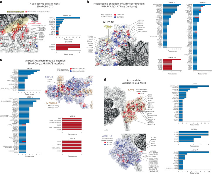
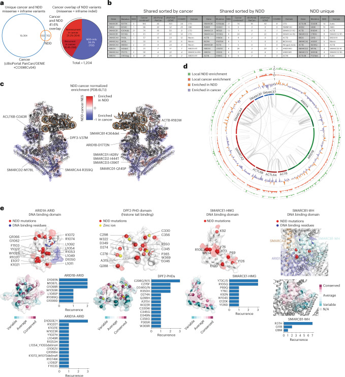
DNA sequencing-based studies of neurodevelopmental disorders (NDDs) have identified a wide range of genetic determinants. However, a comprehensive analysis of these data, in aggregate, has not to date been performed. Here, we find that genes encoding the mammalian SWI/SNF (mSWI/SNF or BAF) family of ATP-dependent chromatin remodeling protein complexes harbor the greatest number of de novo missense and protein-truncating variants among nuclear protein complexes. Non-truncating NDD-associated protein variants predominantly disrupt the cBAF subcomplex and cluster in four key structural regions associated with high disease severity, including mSWI/SNF-nucleosome interfaces, the ATPase-core ARID-armadillo repeat (ARM) module insertion site, the Arp module and DNA-binding domains. Although over 70% of the residues perturbed in NDDs overlap with those mutated in cancer, ~60% of amino acid changes are NDD-specific. These findings provide a foundation to functionally group variants and link complex aberrancies to phenotypic severity, serving as a resource for the chromatin, clinical genetics and neurodevelopment communities.


Conflict of interest statement

C.K. is the scientific founder, scientific advisor to the Board of Directors, scientific advisory board member, shareholder and consultant for Foghorn Therapeutics. C.K. is also a member of the scientific advisory board and is a shareholder of Nested Therapeutics, Nereid Therapeutics and Accent Therapeutics, serves on the scientific advisory board for Fibrogen and serves as a consultant for Google Ventures and Cell Signaling Technologies. C.K. and A.M.V. hold patents in the field of mSWI/SNF complex targeting therapeutics. S.A.S.V. is a member of the scientific advisory board at Ambry Genetics, for which no compensation is received. The other authors declare no competing interests.

Figures

Fig. 1
Fig. 1. Genes encoding chromatin regulatory complexes represent the most frequently mutated gene classes in human NDDs.
a, Scatterplot of the average numbers of non-benign SNVs in DECIPHER corresponding to the top 50 GOMF gene sets enriched in DDG2P developmental disorder-associated genes, ranked by the mutational burden of each gene set. b, Bar graph depicting the total number of NDD-associated missense and protein truncating variants (PTVs) for a curated list of chromatin regulatory and epigenetic gene sets, ranked by mutational burden of each gene set in autism spectrum disorders and developmental disorders (ASD + DD) from the Simons Foundation Research Initiative (SFARI) datasets (SPARK: Simons Foundation Powering Autism Research + SSC-ASC: Simons Searchlight Collection–Autism Sequencing Consortium, and DDD: Deciphering developmental disorders studies). The mSWI/SNF chromatin remodeling complex gene set is emphasized in red. c, Heatmaps depicting the mutational frequency for genes encoding members of the SWI/SNF, CHD, ISWI, and INO80 complex families in DECIPHER. Total number of variants (including copy-number variant (CNV) gain, copy number variant loss (CNV loss), single nucleotide variant (SNV) and indel mutational frequencies are indicated. Percentage of total DECIPHER sequence variants are indicated for each chromatin remodeling complex family (top). d, Cartoon representations of the four classes of chromatin remodelers (SWI/SNF, CHD, ISWI and INO80) and respective subcomplex or related complex associations, colored by CNV loss/SNV/indel variation frequency from panel c. Interchangeable subunit paralogs are colored by their combined mutational frequency. Autism spectrum disorder (ASD) risk score (SFARI) and developmental disorder associations curated from literature and OMIM (Online Mendelian Inheritance in Man, a catalog of human genes and genetic disorders; https://www.omim.org/) are indicated. Asterisk (*) indicates paralog implicated in NDD. Where possible, cartoons were based on 3D structural data available from human and yeast structures; ovals are used in in lieu of structural cartoons for components lacking structural data.
Fig. 2
Fig. 2. Analysis of NDD-associated SNV and indel mutations in mSWI/SNF complex components.
a, Pie chart reflecting the distribution of n = 2,539 mSWI/SNF NDD-associated SNV and in-frame indel mutations from an integrated dataset containing data from SPARK, SSC-ASC, DDD, DECIPHER, ClinVar, LOVD, literature curation and novel variants reported in this study. b, Bar chart summarizing total NDD-associated missense/in-frame deletions and insertions (red) and nonsense/frameshift-inducing mutations (blue) across all mSWI/SNF genes. c, Scatterplot of the negative-normalized ConSurf conservation score versus the mutational recurrence at each mSWI/SNF complex subunit residue for NDD missense and in-frame variants in the integrated dataset. Highly conserved and highly mutated positions are labeled. d, Stacked bar chart summarizing proportion of NDD-associated missense and in-frame insertion/deletion variants falling within (intra, blue) or outside (inter, orange) of mSWI/SNF subunit domains in the integrated dataset. Domains annotated by PFAM, UniProtKB, manual curation, and structurally resolved domains (see also Supplementary Table 3). e, Lollipop plots of NDD mutations in the integrated dataset across protein domain schematics of ARID1A/B, ARID2, SMARCA2/4, SMARCB1, SMARCC1, SMARCE1, and DPF2 subunits generated with Protein Paint. Missense (blue), nonsense (orange), frameshift (red), in-frame deletions (gray) and insertions (brown) are shown. Kernel density estimates (relative frequency distribution) of gnomAD missense mutations (purple line) are overlaid. Domain annotations informed by PFAM, UniProtKB, manual curation, or by structurally resolved domains are indicated. ConSurf conservation scores are shown in a cyan-white-magenta heatmap in increasing conservation order, and structural coverages of the nucleosome core particle (NCP)-bound human cBAF (light orange, PDB: 6LTJ), endogenous human cBAF-NCP bound (red, PDBDEV00000056), and by both structures (brown). Structural coverage for the NCP-bound PBAF complex is also shown for ARID2 (light green, PDB:7VDV). f, PolyPhen HumVar predicted phenotypic severity score and missense mutational recurrence of mSWI/SNF gene mutations from the integrated dataset in intra (blue) and inter (orange) domains.
Fig. 3
Fig. 3. Mapping of 238 unique NDD-associated variant positions onto the structure of the human cBAF complex.
NDD-associated variants, including 14 novel variants, mapped on to the 3D structure of the human cBAF complex (PDB:6LTJ). Residues shown in red spheres represent NDD-associated variants in the subunit indicated, residues in blue represent those mapped from the paralog subunit, and residues in purple represent NDD-variants mapped in both the primary subunit present on the cBAF structure and paralog mapped subunit. Recurrent variants (n ≥ 3) are emphasized in red text. Caution is needed when evaluating these variants in a clinical context since not all variants are confirmed as causal.
Fig. 4
Fig. 4. NDD-associated mutations cluster within key structural hubs of mSWI/SNF complexes.
a, Zoomed-in view of the SMARCB1 C-terminal alpha-helix domain (PDB:6LTJ) with the nucleosome acidic patch interaction site highlighted in yellow (left). NDD-associated mutations in SMARCB1 are emphasized in red. All NDD-associated SMARCB1–C terminal alpha-helix mutations ranked by frequency (right). Novel SMARCB1 variant cases reported in this study shown in red bar chart. b, Zoomed-in view of the SMARCA4 ATPase subunit within the cBAF complex (PDB:6LTJ) at its interface with the nucleosome (left). Mutations in SMARCA4 are indicated in red; mutations in SMARCA2 are indicated in blue, shared mapped in purple. ATP binding pocket is highlighted in yellow. NDD-associated missense and inframeshift variants in SMARCA4 and SMARCA2, ranked by frequency, filtered for recurrence of n ≥ 2 by position (right). Novel SMARCA4 cases reported in this study shown in red bar chart. c, NDD-associated mutations in ARID1A and ARID1B, ranked by frequency, filtered for recurrence of n ≥ 2 by position (left). Zoomed-in view of the SMARCA4-ARID1A interface within the core module of the cBAF complex (right). SMARCA4 is shown in tan and ARID1A in light purple, with mutations in SMARCA4 and ARID1A shown in red and those in their respective paralogs SMARCA2 and ARID1B shown in blue. Novel ARID1A/B variant cases reported in this study shown in red bar chart. d, Left, zoomed-in view of the ACTB (tan) and ACTL6A (light purple) subunits within the Arp module of the cBAF complex, with mutations indicated in red and blue for ACTL6A paralog subunit, ACTL6B. NDD-associated mutations in ACTL6A, ACTL6B and Actin, ranked by frequency, filtered for recurrence of n ≥ 2 by position (right). Recurrent ACTL6B variants donated in brackets mapped onto ACTL6A indicated.
Fig. 5
Fig. 5. Comparison of NDD- and cancer-associated mutations in mSWI/SNF complex components.
a, Venn diagram overlapping unique cancer and NDD missense and in-frame variants (left). Pie chart reflecting breakdown between NDD- and cancer-associated mSWI/SNF missense and in-frame mutations (right). The breakdown of recurrent and non-recurrent cancer variants is shown. b, Top ten recurrent missense and in-frame indel mutations specific to NDD and those shared between NDD and cancer, sorted by frequency in each disease type. Inter- and intradomains are indicated. c, Heatmap representation of mutation differences between NDD and cancer (NDD - Cancer normalized enrichment scores (Methods)) reflected on the 3D structure of the human cBAF complex (PDB:6LTJ). Red regions represent those enriched in NDD, blue represent those enriched in cancer (−1, maximally enriched in cancer; 1, maximally enriched in NDD). Labels for NDD hotspots are shown. d, Circos plot reflecting regions of top-mutated mSWI/SNF subunits and the local enrichment of missense and in-frame indel mutations in NDD (green), Cancer (red) or NDD-Cancer difference (represented as NDD-Cancer NES): NDD (orange) or cancer (purple); interactions between subunits, determined by cross-linking mass-spectrometry (CX-MS) performed on endogenous cBAF complexes are shown (NCP-bound endogenous cBAF, from Mashtalir et al.). Scaled local recurrence, and NDD-Cancer NES were calculated similarly to panel c with one exception, where all secondary paralog mutations were preserved instead of remapping to paralogs. Enrichment scores were bounded from 0 to 1 for local recurrence and −1 to 1 for differential enrichment of mutations. Domains are represented as darker bands in the first inner ring of the Circos plot. e, NDD-associated mutant residues emphasized as red spheres on the structures of the ARID1A-ARID domain (PDB:1RYU), the DPF2-PHD domain (PDB:5B79), the SMARCE1-HMG DNA-binding domain (PDB:7CYU) and the SMARCB1-winged-helix DNA-binding domain (PDB:6LTJ). NDD-associated missense and inframeshift variants, ranked by frequency, are shown as bar charts. ConSurf conservation scores are mapped onto each domain structure with cyan-white-magenta color scale in increasing conservation order.
Fig. 6
Fig. 6. Summary of widely disrupted mSWI/SNF complex hubs in NDDs.
NDD-associated mSWI/SNF mutations occur across several subunits of the mSWI/SNF family of chromatin remodeling complexes and cluster in key structural hubs. Missense and in-frame deletions accumulate within the catalytic ATPase, nucleosome interacting, histone-binding or DNA-binding domains, as well as the ARP module, underscoring their convergence in producing neurodevelopmental aberrations. Interpretation of NDD-associated variants in the context of this framework enables mechanistic dissection of mSWI/SNF activities and provides functional links relevant to clinical phenotypes.
Extended Data Fig. 1
Extended Data Fig. 1. SWI/SNF complex genes are among the most frequently mutated genes in human NDD.
a, Bar charts depicting mean number of non-benign SNVs in DECIPHER and ASD+DD across gene sets indicated. b, Bar graph summarizing the number of non-benign DECIPHER SNVs across top 5 categories from Fig. 1a. c–e, Rank plots depicting GOMF gene sets in (c) DDG2P, (d) ranked by total number of ASD+DD de novo missense variants, (e) ranked by mutation frequency, top 50 GOMFs. f–j, Bar charts showing distribution of variants across sets indicated in each title. mSWI/SNF or cBAF, PBAF, and ncBAF are highlighted in red. k, Heatmap summarizing DECIPHER database mutational frequency for each chromatin remodeling complex separated by variant type (all variants, copy number variants (CNV), and SNVs/indels). l, Pie charts showing inheritance, pathogenicity, and zygosity breakdown of all mSWI/SNF complex variants from DECIPHER. m, Heatmaps depicting the mutational frequency of chromatin remodeling genes in SWI/SNF, CHD, ISWI, and INO80 complex family classes in the ASD+DD dataset. Total number of SNV and indel variants per protein complex family indicated. n, Scatterplot of the total number of de novo missense and PTVs in ASD+DD for all genes ranked by the mutational burden of each gene. mSWI/SNF genes are shown in red. o, Scatterplot of the log normalized total number of cancer missense, frameshift, and nonsense mutations in the TCGA MC3 PanCancer dataset versus the total number of NDD de novo missense and PTVs in ASD+DD datasets. mSWI/SNF genes shown in red. p, Grouped bar graph of the proportion of NDD (blue) and cancer (orange) missense and PTV mutations across all mSWI/SNF genes sorted by decreasing NDD mutational proportion.
Extended Data Fig. 2
Extended Data Fig. 2. Characteristics of NDD-associated single-residue amino acid perturbations in mSWI/SNF components.
a, Distribution of single-nucleotide variants (SNVs) found in NDD-associated missense mutations of mSWI/SNF family genes (Supplementary Table 1) in the integrated dataset (n=2539). b, Horizontal bar graphs of the top 20 amino acid missense substitutions in the integrated dataset (Supplementary Table 1). c, Bar chart characterizing amino acid chemical property changes upon missense mutation for NDD-associated variants in the integrated dataset. d, Stacked bar graphs of the distribution of amino acid substitution chemical property changes in NDD-associated missense mutations in the integrated dataset. e, Sankey diagram of the distribution of NDD-associated missense substitutions in the integrated dataset. Ribbon thickness represents frequency of substitutions in the integrated dataset. f, Stacked bar chart summarizing percentage of NDD-associated missense and in-frame indel mutations in the integrated dataset falling within intrinsically disordered (defined by MobiDB-lite) or structured regions for (left) each mSWI/SNF subunit and (right) all mSWI/SNF subunits combined.
Extended Data Fig. 3
Extended Data Fig. 3. NDD-associated missense variants mapped on cBAF and PBAF 3D structures.
a, NDD-associated missense and inframe indel variants mapped on to the 3D structure of the endogenous human cBAF complex (PDBDEV_00000056). Red spheres represent NDD-associated variants in the subunit indicated, blue spheres represent those mapped from the paralog subunit, and residues in purple represent NDD-variants mapped in both primary subunit present on cBAF structure and paralog subunit. Variants that map exclusively on endogenous complex are indicated. Recurrent variants (n>3) are emphasized in red. b, Bar chart indicating proportion of NDD-associated missense and in-frame indel mutations in the integrated dataset mappable to current mSWI/SNF complex structures separated by subunits. c, NDD-associated missense and inframe indel variants mapped on to the 3D structure of the PBAF complex (PDB 7VDV). Red and blue spheres represent NDD-associated variants in the subunit indicated. Blue spheres and annotations emphasize PBAF subcomplex specific variants mapped.
Extended Data Fig. 4
Extended Data Fig. 4. Structural dissection of mSWI/SNF subunit mutations across the ARP, Core, and ATPase modules.
a, b, (a) SMARCB1-C terminal alpha helix and (b) SMARCA4-ATPase domain (top) ConSurf conservation mapping and (bottom) multiple sequence alignment using D. melanogaster, C. elegans, and S. cerevisiae homologs. c, NDD-associated missense and in-frame indel variants mapped onto the 3D structure of the cBAF complex (PDB:6LTJ) color coded by residue chemical characteristics: red: positive charge, blue: negative charge, green: polar, orange: nonpolar. Nonpolar residues of the ACTB (Arp module) and Table of nonpolar mutations predicted to structurally disrupt ACTB are shown. d, ACTB NDD mutations may alter internal hydrophobic cavities, interfaces with ACTL6A/B, and interfaces with SMARCA2/A4-HSA. Mutant residues shown in red and putative proximal/interacting residues shown in blue/purple. e, SMARCB1-RPT and WH domain NDD mutations predicted to disrupt internal cavity integrity, and hydrogen bonding to interacting ARID1A main chain carbonyls, respectively. Top, selected NDD-associated SMARCB1 missense mutations are labeled, and major domains of SMARCB1 are colored, including RPT1 (blue), RPT2 (orange), and CTD (red). Bottom, mutant residue shown in red and putative proximal/interacting residues shown in blue. f, Mapping of conserved SMARCA2/4 NDD mutant residues (red) on the yeast Snf2 ATPase domain (5X0Y and 6UXW) compared to the recombinant cBAF SMARCA4 ATPase (6LTJ). Brace helices (indicated in yeast structures) are not resolved in human cBAF structure, but demonstrate that certain residues, emphasized in yellow, are buried by the SMARCA2/4 brace helices, rather than exposed. g, Mapping of SMARCA2/4 brace helix NDD variants onto the closed state of the SMARCA4 ATPase domain using the PBAF structure (7VDV). NDD variants clustered in brace helices are predicted to disrupt nucleosome remodeling activity as has been shown with R1243 and R973 NDD and cancer-associated mutations indicated in panel. h, Mapping of SMARCA2/4 NDD mutant residues on the Snf2 ATPase open (gray) and closed (pale cyan) states (PDB IDs: 5Z3O, 5Z3U). NDD residues colored blue in open state and red in closed state. i, SMARCA2/4 NDD mutant residues (left) within 5Å of the ADP-BeFx and (right) interacting with nucleosomal DNA mapped onto the closed yeast Snf2 ATPase structure (5Z3U).
Extended Data Fig. 5
Extended Data Fig. 5. Perturbed subunit positions shared between cancer and NDD highlight ATPase, nucleosome binding regions, and Arp module.
a, Venn diagram overlapping unique cancer missense and inframeshift variants identified from cBioPortal_PanCan, cBioPortal_GENIE and COSMICv94 cancer genetics datasets. b, Venn diagram overlapping unique cancer and NDD (Supplementary Table 1) missense and inframe variants by amino acid position regardless of mutation consequence. NDD mutations derived from Supplementary Table 1, cancer mutations derived by combining cBioPortal_PanCan, cBioPortal_GENIE and COSMICv94 datasets. c, Top ten most recurrent mutant residue amino acid positions shared between Cancer and NDD sorted by frequency in each disease type. Highest recurrence of NDD mutations also included. NDD- and cancer-associated mutations were derived as described in (b). d, Bar plot showing the total number of unique missense/indel mSWI/SNF mutations across the following cancer datasets: cBioPortal_PanCan, cBioPortal_GENIE, COSMICv94. e, Correlation of missense and inframeshift mutations shared between cancer (cBioPortal_PanCan only) and NDD across recombinant cBAF structure. Briefly, NDD- and cancer-associated missense and in-frame indel mutations were remapped onto the primary paralogs of the recombinant cBAF (PDB ID: 6LTJ) structure. A rolling average with a window size of 11aa centered on each residue (5aa on each side) of mutation recurrence by residue position for NDD and cancer was used for the scatterplot and correlation calculation. NDD- and cancer-associated mutations were derived from Supplementary Table 1 (NDD) and cBioPortal_PanCan datasets. The translucent bands around the regression line represent the 95% confidence interval estimated using a bootstrap for 100 iterations. f, Heatmap representation of scaled local enrichment of NDD- and cancer-associated missense and in-frame indel mutational burden of (left, in green) NDD and (right, in red) cancer reflected on the 3D structure of the human cBAF complex (PDB: 6LTJ). Local enrichment scores were computed as described in (Fig. 5e). NDD- and cancer-associated mutations were derived as described in (Fig. 5e). g–j, Multiple sequence alignment of (g) ARID1A-ARID domain, (h) SMARCB1-WH domain, (i) DPF2-PHD domain, and (j) SMARCE1-HMG domain, with variety of related homologs (including M. musculis, D. rerio, D. melanogaster, C. elegans, and S. cerevisiae, where possible).

References

    1. Bailey MH, et al. Comprehensive characterization of cancer driver genes and mutations. Cell. 2018;173:371–385. doi: 10.1016/j.cell.2018.02.060. - DOI - PMC - PubMed
    1. Gabriele M, Tobon AL, D’Agostino G, Testa G. The chromatin basis of neurodevelopmental disorders: Rethinking dysfunction along the molecular and temporal axes. Prog. Neuropsychopharmacol. Biol. Psychiatry. 2018;84:306–327. doi: 10.1016/j.pnpbp.2017.12.013. - DOI - PubMed
    1. Rubeis SD, et al. Synaptic, transcriptional and chromatin genes disrupted in autism. Nature. 2014;515:209–215. doi: 10.1038/nature13772. - DOI - PMC - PubMed
    1. Satterstrom FK, et al. Large-scale exome sequencing study implicates both developmental and functional changes in the neurobiology of autism. Cell. 2020;180:568–584. doi: 10.1016/j.cell.2019.12.036. - DOI - PMC - PubMed
    1. Clapier CR, Iwasa J, Cairns BR, Peterson CL. Mechanisms of action and regulation of ATP-dependent chromatin-remodelling complexes. Nat. Rev. Mol. Cell Biol. 2017;18:407–422. doi: 10.1038/nrm.2017.26. - DOI - PMC - PubMed

Publication types