. 2023 Jul 27;13(1):12147.
doi: 10.1038/s41598-023-39227-x.

High sensitivity of asymmetric 18F-THK5351 PET abnormality in patients with corticobasal syndrome

Affiliations

High sensitivity of asymmetric 18F-THK5351 PET abnormality in patients with corticobasal syndrome

Masanori Kurihara et al. Sci Rep. .

Abstract

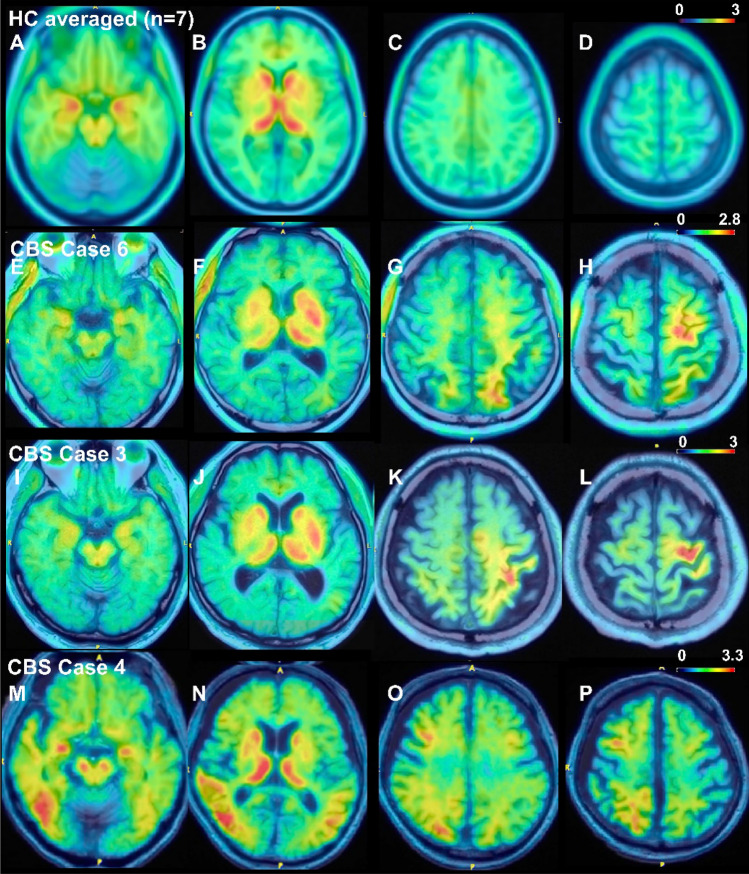
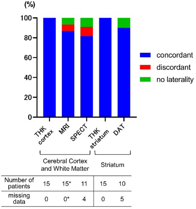
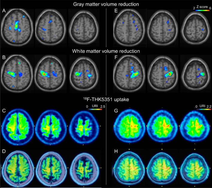
Corticobasal syndrome (CBS) is characterized by symptoms related to the asymmetric involvement of the cerebral cortex and basal ganglia. However, early detection of asymmetric imaging abnormalities can be challenging. Previous studies reported asymmetric 18F-THK5351 PET abnormalities in CBS patients, but the sensitivity for detecting such abnormalities in larger patient samples, including early-stage cases, remains unclear. Patients clinically diagnosed with CBS were recruited. All patients displayed asymmetric symptoms in the cerebral cortex and basal ganglia. Asymmetric THK5351 PET abnormalities were determined through visual assessment. Brain MRI, perfusion SPECT, and dopamine transporter (DAT) SPECT results were retrospectively reviewed. The 15 patients had a median age of 72 years (59-86 years) and a disease duration of 2 years (0.5-7 years). Four patients met the probable and 11 met the possible CBS criteria according to Armstrong criteria at the time of PET examination. All patients, including early-stage cases, exhibited asymmetric tracer uptake contralateral to their symptom-dominant side in the cerebral cortex/subcortical white matter and striatum (100%). The sensitivity for detecting asymmetric imaging abnormalities contralateral to the symptom-dominant side was 86.7% for brain MRI, 81.8% for perfusion SPECT, and 90% for DAT SPECT. White matter volume reduction was observed in the subcortical region of the precentral gyrus with increased THK5351 uptake, occurring significantly more frequently than gray matter volume reduction. THK5351 PET may be a sensitive imaging technique for detecting asymmetric CBS pathologies, including those in early stages.


Conflict of interest statement

The authors declare no competing interests.

Figures

Figure 1
Figure 1
Representative 18F-THK5351 PET image results overlayed on structural brain MRI images. (AD) Average results of seven healthy control volunteers. Physiological tracer uptake was primarily observed in the bilateral striatum, thalamus, and amygdala, where MAO-B is abundant. (EH) Results for a 69-year-old man with possible CBS and a disease duration of one year (Case 6). The patient exhibited ideomotor apraxia, right dominant limb kinetic apraxia, rigidity, dystonia, and myoclonus. CSF biomarkers indicated non-AD. Left-dominated tracer uptake was observed in the frontal and parietal lobes, including the precentral gyrus. (IL) Results for a 70-year-old man with probable CBS and a disease duration of 4 years (Case 3). The patient showed right-dominant limb kinetic apraxia, cortical sensory deficits, mild dysarthria, limb dystonia, and limb myoclonus. Left-dominated tracer uptake was observed in the frontal and parietal lobes, including the precentral and postcentral gyri. (MP) Results of a 71-year-old man with possible CBS and a disease duration of a year (Case 4). The patient showed left-dominant limb kinetic apraxia and limb rigidity. CSF biomarkers suggestive of AD. Right-dominated tracer uptake was observed in the frontal, parietal, and temporal lobes. (EP) Tracer uptake was observed in both the cerebral cortex and associated white matter. Striatal tracer uptake was higher contralateral to the symptom-dominant side in all patients. Color scales represent the uptake ratio index (URI), with the cerebellum as the reference region.
Figure 2
Figure 2
Concordance of imaging abnormality laterality against symptoms. The percentages of patients with asymmetric imaging abnormalities contralateral to the symptom-dominant side (concordant) are shown in blue. *Although brain MRI was available for all 15 patients, volumetric analyses based on 3D T1-weighted images were not available for 5 patients. Volumetric analyses were available for one patient with no laterality (CBS with CSF biomarker suggesting AD) and were unavailable for one patient with discordant results. THK 18F-THK5351 PET, MRI structural magnetic resonance imaging, SPECT brain perfusion single-photon emission tomography, DAT dopamine transporter SPECT.
Figure 3
Figure 3
Representative relation between THK5351 uptake and gray or white matter volume reduction analyses. Imaging results for cases 10 (AD) and 11 (EH). The results of gray (A,E) and white matter (B,F) volume reduction analyses, 18F-THK5351 PET images (C,G), and 18F-THK5351 PET images overlaid on T1-weighted MRI (D,H) are shown. URI uptake ratio index with the cerebellum as the reference region.

References

    1. Boeve BF, Lang AE, Litvan I. Corticobasal degeneration and its relationship to progressive supranuclear palsy and frontotemporal dementia. Ann. Neurol. 2003;54(Suppl 5):S15–S19. doi: 10.1002/ana.10570. - DOI - PubMed
    1. Boeve BF, et al. Pathologic heterogeneity in clinically diagnosed corticobasal degeneration. Neurology. 1999;53:795–800. doi: 10.1212/wnl.53.4.795. - DOI - PubMed
    1. Tartaglia MC, et al. Sporadic corticobasal syndrome due to FTLD-TDP. Acta Neuropathol. 2010;119:365–374. doi: 10.1007/s00401-009-0605-1. - DOI - PMC - PubMed
    1. Josephs KA, et al. Neuropathological background of phenotypical variability in frontotemporal dementia. Acta Neuropathol. 2011;122:137–153. doi: 10.1007/s00401-011-0839-6. - DOI - PMC - PubMed
    1. Dickson DW, et al. Office of Rare Diseases neuropathologic criteria for corticobasal degeneration. J. Neuropathol. Exp. Neurol. 2002;61:935–946. doi: 10.1093/jnen/61.11.935. - DOI - PubMed

Publication types