. 2023 Jul 27;190(8):332.
doi: 10.1007/s00604-023-05888-8.

Uptake and anti-inflammatory effects of liposomal astaxanthin on endothelial cells tracked by Raman and fluorescence imaging

Affiliations

Uptake and anti-inflammatory effects of liposomal astaxanthin on endothelial cells tracked by Raman and fluorescence imaging

Basseem Radwan et al. Mikrochim Acta. .

Abstract

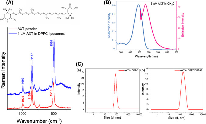
Astaxanthin (AXT) is a lipophilic antioxidant and anti-inflammatory natural pigment whose cellular uptake and bioavailability could be improved via liposomal encapsulation. Endothelial cells (EC) line the lumen of all blood vessels and are tasked with multiple roles toward maintaining cardiovascular homeostasis. Endothelial dysfunction is linked to the development of many diseases and is closely interconnected with oxidative stress and vascular inflammation. The uptake of free and liposomal AXT into EC was investigated using Raman and fluorescence microscopies. AXT was either encapsulated in neutral or cationic liposomes. Enhanced uptake and anti-inflammatory effects of liposomal AXT were observed. The anti-inflammatory effects of liposomal AXT were especially prominent in reducing EC lipid unsaturation, lowering numbers of lipid droplets (LDs), and decreasing intercellular adhesion molecule 1 (ICAM-1) overexpression, which is considered a well-known marker for endothelial inflammation. These findings highlight the benefits of AXT liposomal encapsulation on EC and the applicability of Raman imaging to investigate such effects.

Keywords: Carotenoids; Cellular uptake; Endothelium; Liposomes; Raman microscopy.


Conflict of interest statement

The authors declare no competing interests.

Figures

Fig. 1
Fig. 1
Characterization of astaxanthin-loaded liposomes. A Structure and Raman spectra of AXT powder (red) and in DPPC liposomes (blue). B Absorption and emission spectra of AXT in CH3Cl. C Dynamic light scattering (DLS) results of (a) DPPC liposomes (85 ± 10 nm) and (b) DOTAP-DOPE liposome (185 ± 15 nm), number of experiments (n = 3)
Fig. 2
Fig. 2
EC uptake of AXT-loaded liposomes studied by Raman imaging. Raman images of HAoEC, control, and cells incubated with AXT, AXT-loaded neutral or cationic liposomes, (for 30 min, 1, 3, and 24 h) obtained by the integration of the Raman bands over the selected spectral regions: 3030–2800 cm−1 (C–H stretching), 2900–2830 cm−1 (lipids), 1535–1502 cm−1 (astaxanthin), and composite images showing the distribution of lipids (green), AXT (red), and the overlay in yellow. Raman imaging of all samples was performed once with low laser power (3 mW) to detect AXT Raman bands and once with high laser power (30 mW) to detect bands associated with lipids
Fig. 3
Fig. 3
The effects of free and encapsulated AXT on activated EC lipids studied by Raman imaging. A Raman images of HAoEC, other than the control group; all the presented groups were pre-incubated with TNF-α for 24 h and then incubated with AXT, AXT-loaded  neutral or cationic liposomes, (for 1, 3, or 24 h). Pseudocolor images obtained by the integration of the Raman bands over the selected spectral regions: 2800–3030 cm−1 (C–H stretching), 2830–2900 cm−1 (lipids), 3000–3030 cm−1 (unsaturated lipids), and 1502–1535 cm−1 (AXT). Raman imaging of all samples was performed once with low laser power (3 mW) to detect AXT Raman bands and once with high laser power (30 mW) to detect bands associated with lipids. B Averaged Raman spectra of the lipid class of the control (grey), TNF-α pre-treated group (black), TNF-α pre-treated then incubated with AXT (red), AXT-loaded neutral liposomes (blue), and cationic liposomes (purple). C Quantification of Raman intensity of the band at 3015 cm−1 in different groups. n = 6 cells in each group, 3 independent experiments were performed
Fig. 4
Fig. 4
Fluorescence-based quantification of free and encapsulated AXT anti-inflammatory effects. A ICAM-1 expression and B lipid droplets (LDs) per cell of control and TNF-α pre-treated groups that were later treated with AXT, AXT-loaded neutral liposomes, or AXT-loaded cationic liposomes. The results are presented as the means + SD from 3 independent experiments, * P < 0.05, ** P < 0.01, and *** P < 0.001. C Representative fluorescence images of HAoEC, showing nuclei (Hoechst stained, blue), lipids (BODIPY stained, green), and ICAM-1 (red). Scale bars equal 50 μm

References

    1. Akbarzadeh A, Rezaei-Sadabady R, Davaran S, et al. Liposome: classification, preparation, and applications. Nanoscale Res Lett. 2013;8:102. doi: 10.1186/1556-276X-8-102. - DOI - PMC - PubMed
    1. Trif M, Craciunescu O. Nanotechnology and functional foods. Chichester, UK: John Wiley & Sons, Ltd; 2015. Liposome as efficient system for intracellular delivery of bioactive molecules; pp. 191–213.
    1. Pereira C, Souza A, Vasconcelos A, et al. Antioxidant and anti-inflammatory mechanisms of action of astaxanthin in cardiovascular diseases (Review) Int J Mol Med. 2020;47:37–48. doi: 10.3892/ijmm.2020.4783. - DOI - PMC - PubMed
    1. Rao AR, Sindhuja HN, Dharmesh SM, et al. Effective inhibition of skin cancer, tyrosinase, and antioxidative properties by astaxanthin and astaxanthin esters from the green alga Haematococcus pluvialis. J Agric Food Chem. 2013;61:3842–3851. doi: 10.1021/jf304609j. - DOI - PubMed
    1. Visioli F, Artaria C. Astaxanthin in cardiovascular health and disease: mechanisms of action, therapeutic merits, and knowledge gaps. Food Funct. 2017;8:39–63. doi: 10.1039/C6FO01721E. - DOI - PubMed

Publication types

LinkOut - more resources