Review
. 2023 Sep;483(3):349-365.
doi: 10.1007/s00428-023-03607-5. Epub 2023 Jul 27.

Follicular helper T-cell lymphomas: disease spectrum, relationship with clonal hematopoiesis, and mimics. A report of the 2022 EA4HP/SH lymphoma workshop

Affiliations
Review

Follicular helper T-cell lymphomas: disease spectrum, relationship with clonal hematopoiesis, and mimics. A report of the 2022 EA4HP/SH lymphoma workshop

Sarah L Ondrejka et al. Virchows Arch. 2023 Sep.

Abstract

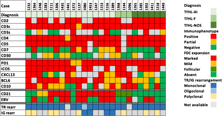
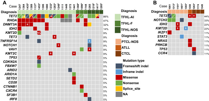
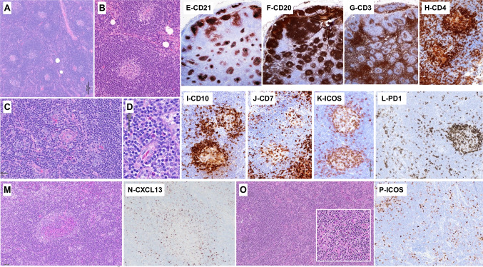
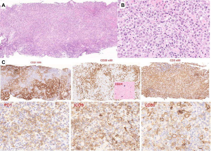
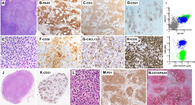
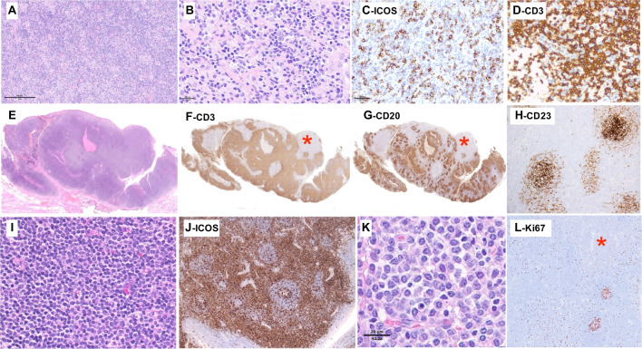
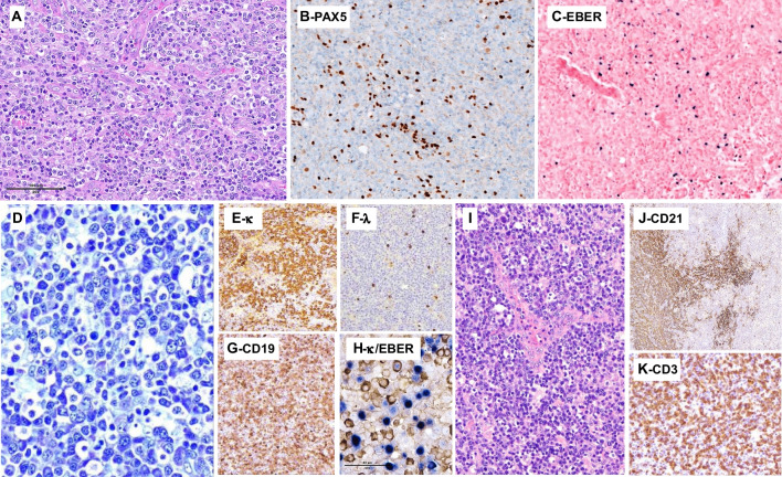
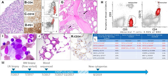
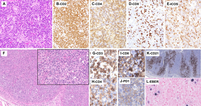
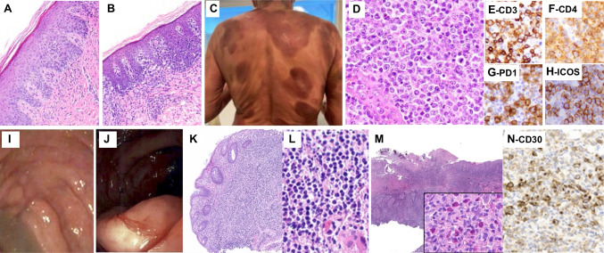
Follicular helper T-cell lymphomas (TFH lymphomas) were discussed in session V of the lymphoma workshop of the European Association for Haematopathology (EA4HP)/Society for Hematopathology (SH) 2022 meeting in Florence, Italy. The session focused on the morphologic spectrum of TFH lymphoma, including its three subtypes: angioimmunoblastic-type (AITL), follicular-type, and not otherwise specified (NOS). The submitted cases encompassed classic examples of TFH lymphoma and unusual cases such as those with early or indolent presentations, associated B-cell proliferations, or Hodgkin/Reed-Sternberg-like cells. The relationship between TFH lymphoma and clonal hematopoiesis was highlighted by several cases documenting divergent evolution of myeloid neoplasm and AITL from shared clonal mutations. The distinction between TFH lymphoma and peripheral T-cell lymphoma, not otherwise specified (PTCL, NOS), was stressed, and many challenging examples were presented. Various cases highlighted the difficulties of differentiating TFH lymphoma from other established types of lymphoma and reactive conditions. Cutaneous T-cell lymphoma expressing TFH markers, particularly when resulting in lymph node involvement, should be distinguished from TFH lymphomas. Additional immunophenotyping and next-generation sequencing studies were performed on various cases in this session, highlighting the importance of these technologies to our current understanding and classification of TFH lymphomas.

Keywords: Angioimmunoblastic T-cell lymphoma; Clonal hematopoiesis; EA4HP workshop; High-throughput sequencing; Peripheral T-cell lymphoma; T-cell lymphoma; TFH lymphoma.


Conflict of interest statement

The authors declare no competing interests.

Figures

Fig. 1
Fig. 1
Heatmap representation of the immunophenotype, EBV, and clonality results in the 25 follicular helper T-cell lymphomas (TFHLs) of session V. By immunohistochemistry, the neoplastic T cells were positive for CD2 (18/18), cCD3 (25/25, dim in one case), CD4 (24/24, dim in one case), and CD5 (23/23). The aberrant CD3-/dim CD4+ population reported as a common feature of TFH lymphoma [43] was confirmed in this cohort, as 8/12 cases analyzed by flow cytometry were negative or dim for sCD3 expression. Dim, partial, or negative CD7 expression was found in 17/23 cases. Most cases (17/25), including virtually all of those analyzed by flow cytometry (11/12) showed an aberrant T-cell phenotype regarding pan T-cell antigen expression. Some degree of CD30 positivity in the neoplastic cells was reported in 8/19 cases. Considering the five TFH markers currently recommended as a systematic IHC panel [15, 16], PD1 and ICOS were tested in most cases—25 and 18 cases, respectively—and were usually strongly expressed. The expression of CXCL13, BCL6, and CD10 was more heterogeneous. CXCL13 was interpreted as negative in 4/18 cases, partially positive in 6, and strongly positive in 8 cases. BCL6 expression was found in 20/22 cases but was often partial or weak. CD10 was the least sensitive TFH marker, positive in 17/23 cases, but only weak or partial in 7 cases. Only 50% of the cases in this series, including 9/16 AITL, contained EBV-positive B-blasts. A monoclonal T-cell receptor gene (TR gene) rearrangement was demonstrated in 22/23 cases tested, and the remaining case had an oligoclonal TR gene rearrangement. Four of 11 cases tested for an IG gene rearrangement had a B-cell clone
Fig. 2
Fig. 2
Summary of mutated genes in A 22 patients with follicular helper T-cell lymphoma and B 8 patients with other T-cell lymphoma types. Genes mutated in at least two patients and/or with one probably pathogenic (ACGS class 4) or pathogenic (ACGS class 5) mutation are shown. For each gene, the percentage of patients carrying mutation(s) is indicated to the right of the table. High-throughput sequencing results were provided by the submitters, or generated by the panel (*, see supplementary information). ǂ denotes ACGS class 3 mutations (variants of unknown significance). Of note, the assays used for genetic profiling did not assess potential gene fusions, for example, those involving CD28, ICOS, and VAV1, which represent other oncopathogenic mechanisms in TFHL [13, 91]
Fig. 3
Fig. 3
AITL with partial/subtle lymph node (LN) involvement. LYWS-1362 (Pedro Farinha): at low-power view (A), the LN presents a globally preserved architecture, with numerous follicles separated by interfollicular areas; (B) the follicles have small or regressive GCs and thick mantle zones; (C) on close inspection of the interfollicular areas, there is focal increased vascularity and aggregates of slightly atypical cells, also shown in (D); panoramic views of the immunostains (EG) show CD21+ follicular dendritic cell meshworks confined to follicles (E), numerous CD20+ follicles and few extrafollicular B-cells (F), while CD3+ cells constitute the majority of extrafollicular cells but also are increased at the periphery of the GCs (G); the numerous T cells present at the periphery of the GCs and the inner zone of the mantle zone (HL) are positive for CD4 (H) strongly CD10+ (I), negative for CD7 (J), positive for ICOS (K), and positive for PD-1 (L); note that PD-1 also highlights clusters of extrafollicular cells. LYWS-1248 (Danielle Maracaja): this LN that had a globally preserved architecture contains aggregates of atypical clear cells, distributed at the periphery of the GCs and outside follicles (M), and these cells are highlighted by CXCL13 (N). LYWS-1283 (Mats Ehinger): this medium magnification of the HE slide shows a follicle (left) with preserved mantle zone, and mildly expanded interfollicular area, containing increased vessels and an atypical cellular infiltrate (inset) (O); ICOS highlights cells distributed at the periphery of GCs (upper left) as well as in extrafollicular areas (P)
Fig. 4
Fig. 4
AITL diagnosed on a core needle biopsy. LYWS-1441 (Pierre Isnard): wide LN core needle biopsy, showing an effaced architecture, increased vascularity (A) and an atypical cellular infiltrate of clear cells, admixed with small lymphocytes and histiocytes (B); on immunostains (C), CD21 highlights an expanded meshwork of follicular dendritic cells distributed around increased vessels, CD20 stains residual aggregates of small B-cells, as well as scattered larger B-cells, which are positive for EBV by in situ hybridization (inset), the majority of the infiltrate consists of CD3+ cells, which show extensive expression of PD-1 and ICOS, and CD30 stains the large blastic cells as well as a proportion of the atypical T-cells
Fig. 5
Fig. 5
Follicular helper T-cell lymphoma, follicular-type. LYWS-1368 (Emily Mason): in this case, resembling progressive transformation of the GCs, low-power view of the LN shows large nodules, comprising pale cellular aggregates (A); PAX5 shows high B-cell content in the nodules, with a moth-eaten appearance (B), and CD3 stains the intermixed aggregates of clear cells intermixed within the nodules (C); CD21 highlights follicular dendritic cells meshworks underlining the nodules (D); on high-power view (EH), the pale aggregates comprise atypical medium-sized cells with abundant cytoplasm, and scattered blastic cells (E), CD30 stains strongly the large blastic cells (which are also CD20+, not shown) and moderately the atypical clear cells (F); the latter shows positive expression of CXCL13 (G) and ICOS (H); flow cytometry (I) demonstrates a population of T cells with an aberrant immunophenotype (sCD3-, CD5+, and CD7+; green dots). LYWS-1096 (Stefano Lazzi): in this follicular lymphoma-like case, a low panoramic view of the LN (J) shows a vaguely nodular pattern, which is confirmed by CD21 immunostaining (K); the nodules comprised a polymorphic infiltrate including small to medium size atypical cells, large scattered blasts, sometimes resembling Reed–Sternberg cells, and eosinophils (L); PD-1 stains the majority of cells in a nodular pattern (M); on double immunostaining with CD10 (red) and PAX-5 (brown), the nodules comprised PAX5- CD10+ cells (N)
Fig. 6
Fig. 6
Follicular helper T-cell lymphoma, NOS. LYWS-1302 (Wendy Lim Wen-Hsuan): this LN comprises an epithelioid cell-rich infiltrate (A), including atypical lymphoid cells, sometimes with clear cytoplasm (B); the atypical cells are positive for ICOS (C); and the majority were CD3+ (D). LYWS-1218 (Marco Pizzi) in this tonsillectomy specimen, panoramic views (EG) show expanded lymphoid tissue, including a marked expansion of CD3+ areas (F) and preserved CD20+ follicles (G); note the presence of a focus (*) negative for both CD3 and CD20; CD23 immunostaining (H) showed follicular dendritic cells restricted to follicles; the T-cell zones (I, J) comprised a monotonous infiltrate of small to medium-sized lymphoid cells, showing slight atypia being strongly positive for several TFH markers including ICOS shown here (J); the area negative for CD20 and CD3 (*) corresponded to sheets of mature plasma-cells (K); Ki67 shows very low proliferation index, both in the T-cell zones and in the plasma cells infiltrate (*)(L)
Fig. 7
Fig. 7
B-cell proliferations in TFH lymphoma. LYWS-1137 (Nives Zimmermann): this TFHL occurring in a particularly young patient (30-year-old) shows a typical morphology of AITL, including prominent arborising blood vessels, and prominent clear cells (note that this case harboured an IDH2 mutation) (A); PAX5 highlighted a moderate number of large nuclei (B) that were positive for EBER by in situ hybridization (C). LYWS-1080 (Katrin Kurz): this LN comprises a prominent lymphoplasmacytic infiltrate (D) containing many plasmacytic cells monotypic for kappa (E) and with only very few lambda-positive cells (F), as well as numerous B cells highlighted by CD19 (G); on combined staining for kappa and EBER (H), there are many EBV+ cells that mostly correspond to the B cells, while most kappa+ plasma cells were EBER-negative (H); the LN also comprises prominent arborising vessels (I) and CD21 highlights expanded follicular dendritic cells (J); CD3 reveals a diffuse T-cells infiltrate (K) positive for several TFH markers (not shown)
Fig. 8
Fig. 8
Divergent evolution of clonal hematopoiesis to angioimmunoblastic T-cell lymphoma and therapy-related myeloid neoplasm. LYWS-1247 (Natasha Lewis): 72-year-old male diagnosed with AITL on core needle biopsy (A), positive for CD4 (B), CD10 (C), and PD1 (D) by immunohistochemistry and with surface loss of CD3 by flow cytometry (E, F). A staging bone marrow biopsy demonstrated involvement (arrows, G) and bulk peripheral blood flow cytometry showed an exceedingly low level of involvement (H). Two years later, a bone marrow biopsy demonstrated dysmegakaryopoiesis (I), with hypocellular bone marrow (J) and increased blasts (CD34, K). Two TET2 mutations and a DNMT3A were shared between the staging bone marrow involved by AITL, the bulk peripheral blood at diagnosis, and bulk bone marrow two years later, but only the specimen with morphologic AITL involvement demonstrated the RHOA p.G17V and IDH2 p.R172K, and the bone marrow with myeloid progression acquired additional mutations (L). Timeline of events (M)
Fig. 9
Fig. 9
Peripheral T-cell lymphoma, NOS with overlapping TFH features. (Upper row) LYWS-1061 (Megan Fitzpatrick): 68-year-old male with three months of B symptoms, splenomegaly, and stage III lymphadenopathy. H&E shows diffuse small- to medium-sized abnormal lymphocytes and epithelioid histiocytes with occasional giant cells, imparting a Lennert-like appearance. (A) The lymphoma cells are positive for CD2 (B) and CD4 (C), negative for CD8 (D), and have partial expression of ICOS (E). Other TFH markers were negative. NGS identified two TET2 variants and NOTCH2 p.Gln2285Ter (c.6853C>T). There were insufficient TFH markers for a diagnosis of TFH lymphoma. (Lower row) LYWS-1401 (Anne-Roos Schrader): 44-year-old male with stage III lymphadenopathy, and splenic and tonsillar involvement by lymphoma. H&E shows a polymorphous infiltrate with intermediate-sized abnormal lymphocytes, eosinophils, and arborizing blood vessels, imparting an AITL-like appearance (F). Lymphoma cells are positive for CD3 (G). CD4 stains background smaller T-cells and histiocytes (H). CD8 is positive in the atypical lymphocytes (I), and there is partial PD1 coexpression (J). ICOS and BCL6 were also positive. CD21 demonstrated expanded and proliferated FDC meshworks (K). EBER-CISH showed scattered positivity (L)
Fig. 10
Fig. 10
Cutaneous T-cell lymphoma and reactive lymphoproliferations can express TFH markers. (Upper row) LYWS-1174 (Yi Zhou): 78-year-old female diagnosed with cutaneous T-cell lymphoma 4 years ago (A) with worsening skin lesions (B, C), and generalized lymphadenopathy. LN biopsy demonstrated a diffuse proliferation of large neoplastic lymphocytes (D), positive for CD3 (E), CD4 (F), PD1 (G), ICOS (H), CD10 (subset), BCL6 (subset), and negative for CD7. A clonal TR gene rearrangement was identified. A TP63::ARID5B fusion was identified. No mutations in RHOA, IDH2, DNMT3A, or TET2 were detected. (Lower row) LYWS-1197 (Philip Butlerys): intestinal TFH lymphoproliferation in a patient with activated phosphoinositide 3-kinase delta syndrome in a 14-year-old girl on IVIG with recurrent infections, chronic stable lymphadenopathy, and abdominal pain and diarrhea. Colonoscopy revealed areas of nodularity (I) and inflammation with ulceration (J). Histologically, multiple areas of the bowel showed a monotonous small lymphoid infiltrate (K, L) positive for CD3, PD1 (subset), and ICOS. LYWS-1212 (Carlo Pescia): a CD30-positive lymphoproliferation consistent with TUGSE in a 38-year-old HIV-negative male with sudden onset of oral aphthous ulcerations (M), composed of abnormally enlarged lymphocytes with associated eosinophils (inset). Many T-cells were positive for CD3, CD4, and PD1 and negative for cytotoxic markers and EBV. A clonal TR gene rearrangement was identified. CD30 (N) is positive in many of the lymphocytes, which has been reported in cases of TUGSE

References

    1. Mintz MA, Cyster JG. T follicular helper cells in germinal center B cell selection and lymphomagenesis. Immunol Rev. 2020;296(1):48–61. - PMC - PubMed
    1. Grogg KL, Attygalle AD, Macon WR, Remstein ED, Kurtin PJ, Dogan A. Expression of CXCL13, a chemokine highly upregulated in germinal center T-helper cells, distinguishes angioimmunoblastic T-cell lymphoma from peripheral T-cell lymphoma, unspecified. Mod Pathol. 2006;19(8):1101–1107. - PubMed
    1. de Leval L, Rickman DS, Thielen C, Reynies A, Huang YL, Delsol G, et al. The gene expression profile of nodal peripheral T-cell lymphoma demonstrates a molecular link between angioimmunoblastic T-cell lymphoma (AITL) and follicular helper T (TFH) cells. Blood. 2007;109(11):4952–4963. - PubMed
    1. Piccaluga PP, Agostinelli C, Califano A, Carbone A, Fantoni L, Ferrari S, et al. Gene expression analysis of angioimmunoblastic lymphoma indicates derivation from T follicular helper cells and vascular endothelial growth factor deregulation. Cancer Res. 2007;67(22):10703–10710. - PubMed
    1. Swerdlow SH, Campo E, Harris NL, et al. WHO classification of tumours of haematopoietic and lymphoid tissues. Lyon, France: IARC Press; 2008.

MeSH terms