. 2023 Aug;22(8):1039-1046.
doi: 10.1038/s41563-023-01611-3. Epub 2023 Jul 27.

Fibre-infused gel scaffolds guide cardiomyocyte alignment in 3D-printed ventricles

Affiliations

Fibre-infused gel scaffolds guide cardiomyocyte alignment in 3D-printed ventricles

Suji Choi et al. Nat Mater. 2023 Aug.

Abstract

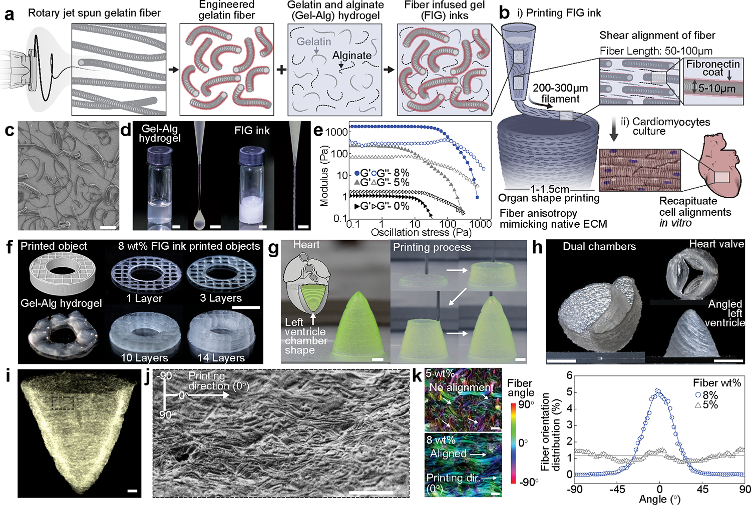
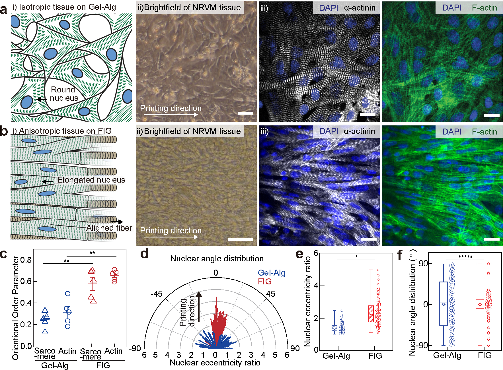
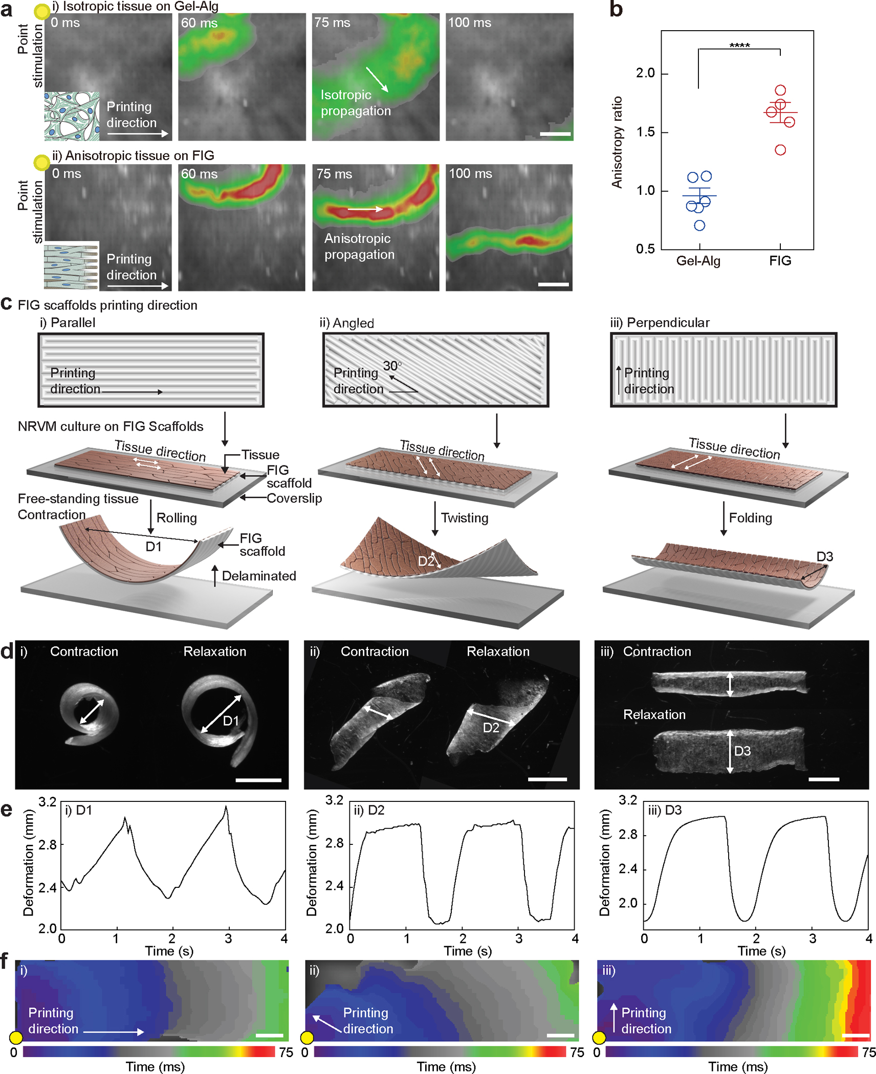
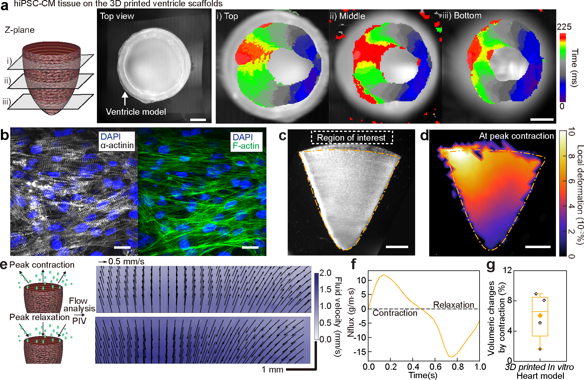
Hydrogels are attractive materials for tissue engineering, but efforts to date have shown limited ability to produce the microstructural features necessary to promote cellular self-organization into hierarchical three-dimensional (3D) organ models. Here we develop a hydrogel ink containing prefabricated gelatin fibres to print 3D organ-level scaffolds that recapitulate the intra- and intercellular organization of the heart. The addition of prefabricated gelatin fibres to hydrogels enables the tailoring of the ink rheology, allowing for a controlled sol-gel transition to achieve precise printing of free-standing 3D structures without additional supporting materials. Shear-induced alignment of fibres during ink extrusion provides microscale geometric cues that promote the self-organization of cultured human cardiomyocytes into anisotropic muscular tissues in vitro. The resulting 3D-printed ventricle in vitro model exhibited biomimetic anisotropic electrophysiological and contractile properties.


Conflict of interest statement

Competing interests

Harvard University filed for intellectual property relevant to this manuscript, listing L.A.M. and K.K.P. as inventors. The remaining authors declare no competing interests.

Figures

Fig. 1.
Fig. 1.. Development of gelatin fiber infused gel (FIG) inks for free-standing 3D printed tissue scaffolds with cellular alignment cues.
a-b, Schematic illustration of FIG ink components. As fragmented gelatin fibers are combined with Gel-Alg hydrogels (Red, Fibronectin (FN)) (a), the ink viscosity increases and displays a solid-like behavior, which allows for 3D printing of ventricle scaffolds with hierarchical structure (b). Fiber alignment occurred under shear stress (τ) during 3D printing leads to native ECM anisotropic structural features in 3D scaffolds, promoting tissue alignment and organization to recapitulate in vivo heart muscle (b). c, A scanning electron microscopy image showing truncated gelatin fibers. Scale bar, 100 μm. d, Comparison of Gel-Alg ink (0 wt% fiber, left panel) or FIG ink (8 wt% fiber, right panel). The Gel-Alg hydrogel has low viscosity liquid-like behavior (left). FIG inks behave solid-like at rest and extrude in a continuous stream (right). Scale bar, 2mm. e, Oscillation stress sweep test to measure storage (Gʹ) and loss (Gʺ) modulus, showing concentration- and strain-dependent shear thinning behavior and sol-gel transition f, 3D donut shape with rectilinear infill pattern with increasing number of stacking layers, showing high shape retention of using FIG inks. g, A cone-shaped model of the self-supporting inverted left ventricle printed in circumferential direction. Scale bars, 2 mm. h, Self-supportive dual ventricle chambers and a heart valve printed in circumferential direction and an angled left ventricle printed in diagonal (30°) titled direction. Scale bars, 5 mm. i, A micro-computed tomography image of the 3D printed ventricle scaffold after critical point drying, showing fiber structure in the 3D printed geometry. Scale bars, 1 mm. j, A scanning electron microscopy image of the 3D printed ventricle scaffold showing fiber alignments in printing direction. Scale bar, 200 μm. k, Analysis of fiber alignment from confocal images of the 3D printed ventricle FIG scaffolds with 5 and 8 wt% fibers and corresponding fiber orientation angular distribution graph. 0° indicates printing direction. Scale bars, 100 μm.
Fig. 2.
Fig. 2.. Anisotropic intra- and inter-cellular organization of cardiac tissues cultured on printed FIG scaffolds.
a-b, i) Schematic illustrations showing neonatal rat ventricular myocyte (NRVM) tissue formation on the 2D printed Gel-Alg hydrogel (a) and FIG (b) scaffolds, ii) brightfield images of NRVM cultured on each scaffold, and iii) representative immunostained images of nuclei (blue), α-actinin (grey), and F-actin (green). Scale bars, 20 μm. c, Normalized sarcomeric α-actinin and F-actin alignment with their alignment quantified on a scale of 0 (random) to 1 (aligned) using an orientation order parameter. Statistical analysis was performed using a two-tailed student’s t-test with unequal variance, **P=0.000252 and 0.00149 for sarcomere and F-actin, respectively. n=5 tissues per scaffold condition. Data are presented as mean values +/− SEM. d, Representative distribution of nuclear shape (line length: eccentricity ratio) and orientation (line angle) with printing direction at 0°. e-f, Nuclear eccentricity ratio (e) and angle (f) from −90° to 90° (n = 203 and 183 nuclei from 3 tissues on Gel-Alg and FIG scaffolds, respectively). Statistical analysis was performed using a two-tailed student’s t-test with unequal variance, *P=0.0295 (e) and a two-sample Kolmogorov-Smirnov test, *****P=3.46e-10. (f). n =203 and 183 nuclei from 3 independent tissues on Gel-Alg and FIG scaffolds, respectively. Box plot: center diamond, box limits, and whiskers indicates mean, the first and third quartiles, and max-min, respectively.
Fig. 3.
Fig. 3.. Dynamics of electromechanical coupling of multi-directional anisotropic cardiac tissues.
a, Representative Ca2+ transient propagation images from Ca2+ optical mapping of NRVM tissues cultured on Gel-Alg scaffolds (i) and FIG scaffolds (ii), resulting in isotropic (i) and anisotropic (ii) Ca2+ propagation, respectively, under 1 Hz point electrical stimulation at the top-left corner (yellow dot). Scale bar, 2 mm. b, The anisotropy ratio (Vlong/Vtrans) for NRVM tissues on Gel-Alg scaffolds and FIG scaffolds, quantifying anisotropic Ca2+ propagation. Statistical analysis was performed using a two-tailed student’s t-test with unequal variance. ****P = 0.000174. n=6,5 tissues per scaffold condition. Data are presented as mean values +/− SEM. c,d, Schematic illustration (c) and microscope images (d) of NRVM tissue cultured on rectangular shaped FIG scaffolds printed in parallel (i), angled (ii), and perpendicular (iii) direction to the long side of scaffold geometry. Delaminated free-standing tissue layers from the coverslip showed different contractile motions, rolling (i), twisting (ii), and bending (iii). Scale bar, 2mm. e, Diameter (arrow in (d)) of the deformed shape continuously changes in time under 0.5 Hz field electrical stimulation. f, Isochrone mapping images demonstrating Ca2+ propagation of NRVM tissues on parallel (i), angled (ii), and perpendicular (iii) patterned FIG scaffolds. Point electrical stimulation at 1 Hz (yellow dot) initiates Ca2+ propagation, showing fast propagation in parallel and angled patterns but slow propagation in the perpendicular pattern throughout the scaffold geometry. Scale bar, 1 mm
Fig. 4.
Fig. 4.. Structural, electrophysiological, and contractile properties of human stem cell-based tissue-engineered 3D ventricle models.
a, Spontaneous Ca2+ transient propagation on a human induced pluripotent stem cell-derived cardiomyocyte (hiPSC-CM) 3D printed ventricle model (top view), showing anisotropic Ca2+ propagation along the printing direction. Scale bar, 2 mm. b, Immunostained images of hiPSC-CM tissues cultured in 3D printed model ventricle scaffolds. Scale bar, 20 μm. c, A brightfield image of the tissue-engineered 3D ventricle model after hiPSC-CMs were cultured for 14 days. Scale bar, 2 mm. d, Representative deformation map of the 3D hiPSC-CM ventricle model at peak contraction. Scale bar, 2 mm. e, Fluid velocity maps at peak contraction (top) and peak relaxation (bottom) analyzed by tracking fluorescent bead displacement in a region of interest under 1 Hz electrical field stimulation. Scale bar, 1 mm. f, Instantaneous mass flux in a region of interest for a representative ventricle during a complete cycle of contraction and relaxation. g, Volumeric changes by contraction of hiPSC-CM 3D ventricle models. n = 4 ventricle models. Box plot: center diamond, box limits, and whiskers indicates mean, the first and third quartiles, and max-min, respectively.

Similar articles

Cited by

References

    1. Frantz C, Stewart KM, Weaver VM The extracellular matrix at a glance. J. Cell Sci. 123, 4195–4200 (2010) - PMC - PubMed
    1. Blazeski A, Lowenthal J, Zhu R, Ewoldt J, Boheler KR, Tung L Functional properties of engineered heart slices incorporating human induced pluripotent stem cell-derived cardiomyocytes. Stem Cell Reports 12, 982–995 (2019) - PMC - PubMed
    1. LeGrice I, Pope A, Smaill B The architecture of the heart: Myocyte organization and the cardiac extracellular matrix. In: Villarreal FJ (ed). Interstitial fibrosis in heart failure. Springer New York: New York, NY, 2005, pp 3–21.
    1. Ronaldson-Bouchard K, Ma SP, Yeager K, Chen T, Song L, Sirabella D, et al. Advanced maturation of human cardiac tissue grown from pluripotent stem cells. Nature 556, 239–243 (2018) - PMC - PubMed
    1. Kleber AG, Rudy Y Basic mechanisms of cardiac impulse propagation and associated arrhythmias. Physiol. Rev. 84, 431–488 (2004) - PubMed

Methods-only references

    1. Rezakhaniha R, Agianniotis A, Schrauwen JT, Griffa A, Sage D, Bouten CV, et al. Experimental investigation of collagen waviness and orientation in the arterial adventitia using confocal laser scanning microscopy. Biomech. Model. Mechanobiol. 11, 461–473 (2012) - PubMed
    1. Lian X, Zhang J, Azarin SM, Zhu K, Hazeltine LB, Bao X, et al. Directed cardiomyocyte differentiation from human pluripotent stem cells by modulating wnt/beta-catenin signaling under fully defined conditions. Nat. Protoc. 8, 162–175 (2013) - PMC - PubMed
    1. Al Tanoury Z, Zimmerman JF, Rao J, Sieiro D, McNamara HM, Cherrier T, et al. Prednisolone rescues duchenne muscular dystrophy phenotypes in human pluripotent stem cell-derived skeletal muscle in vitro. Proc. Natl. Acad. Sci. U. S. A. 118, (2021) - PMC - PubMed
    1. Alex Liberzon DL, Aubert Mathias, Bachant Pete, jakirkham, ranleu, tomerast, Käufer Theo, Borg Joe, Dallas Cameron, & Vodenicharski Boyko. Openpiv/openpiv-python: Fixed windows conda-forge failure with encoding. 10.5281/zenodo.3566451 (2019) - DOI
    1. Choi S, Lee KY, Kim SL, Macqueen LA, Chang H, Zimmerman JF, Jin Q, Peters MM, Ardoña HAM, Liu X, Heiler A-C, Richardson C, Gabardi R, Pu WT, Bausch AR, and Parker KK Fiber Infused Gel Scaffolds Guide Cardiomyocyte Alignment in 3D Printed Ventricles, Figshare, 10.6084/m9.figshare.22787714, 2023 - DOI - PMC - PubMed

Publication types