. 2023 Jul 27;24(1):422.
doi: 10.1186/s12864-023-09505-z.

Diagnostic yield and novel candidate genes for neurodevelopmental disorders by exome sequencing in an unselected cohort with microcephaly

Affiliations

Diagnostic yield and novel candidate genes for neurodevelopmental disorders by exome sequencing in an unselected cohort with microcephaly

Chunli Wang et al. BMC Genomics. .

Abstract

Objectives: Microcephaly is caused by reduced brain volume and most usually associated with a variety of neurodevelopmental disorders (NDDs). To provide an overview of the diagnostic yield of whole exome sequencing (WES) and promote novel candidates in genetically unsolved families, we studied the clinical and genetic landscape of an unselected Chinese cohort of patients with microcephaly.

Methods: We performed WES in an unselected cohort of 103 NDDs patients with microcephaly as one of the features. Full evaluation of potential novel candidate genes was applied in genetically undiagnosed families. Functional validations of selected variants were conducted in cultured cells. To augment the discovery of novel candidates, we queried our genomic sequencing data repository for additional likely disease-causing variants in the identified candidate genes.

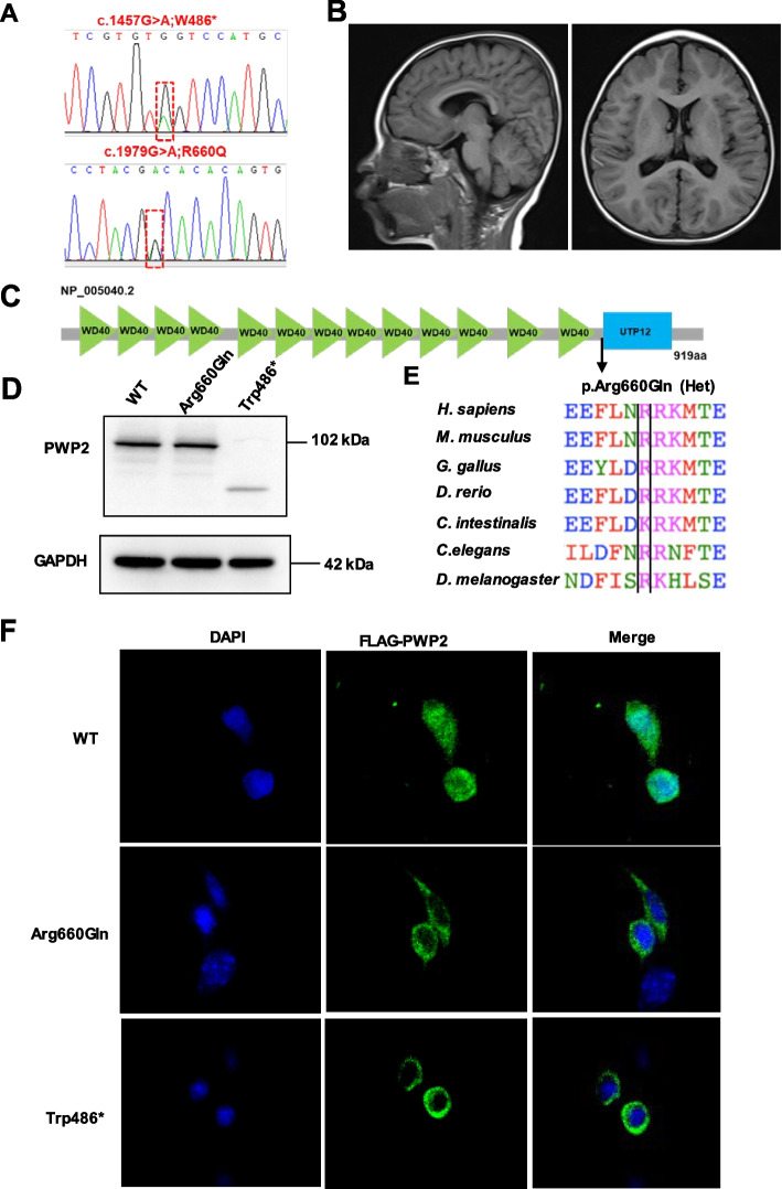
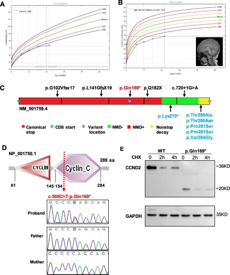
Results: In 65 families (63.1%), causative sequence variants (SVs) and clinically relevant copy number variants (CNVs) with a pathogenic or likely pathogenic (P/LP) level were identified. By incorporating coverage analysis to WES, a pathogenic or likely pathogenic CNV was detected in 15 families (16/103, 15.5%). In another eight families (8/103, 7.8%), we identified variants in newly reported gene (CCND2) and potential novel neurodevelopmental disorders /microcephaly candidate genes, which involved in cell cycle and division (PWP2, CCND2), CDC42/RAC signaling related actin cytoskeletal organization (DOCK9, RHOF), neurogenesis (ELAVL3, PPP1R9B, KCNH3) and transcription regulation (IRF2BP1). By looking into our data repository of 5066 families with NDDs, we identified additional two cases with variants in DOCK9 and PPP1R9B, respectively.

Conclusion: Our results expand the morbid genome of monogenic neurodevelopmental disorders and support the adoption of WES as a first-tier test for individuals with microcephaly.

Keywords: Microcephaly; Neurodevelopmental disorders; Novel candidates; Whole exome sequencing.


Conflict of interest statement

The authors declare no competing interests.

Figures

Fig. 1
Fig. 1
Molecular diagnostic yield and monogenic variants inheritance in 103 families with primary or secondary microcephaly. A, Number and percentage of 103 patients in which P/LP level variants in a monogenic cause (49/103, 47.6%), VUS level variants in a monogenic cause (6/103, 5.8%), a known pathogenic CNV (15.5%) or a newly reported or potential new candidate gene (7.8%) was detected by whole-exome sequencing. Blue color denotes that a variant in genes with established disease phenotypes in humans was detected. Green color denotes that a pathogenic or likely pathogenic CNV was detected. Red was chosen if one highly potential novel gene was detected in the family. Yellow indicates no causative or candidate variants were detected. VUS, variant of uncertain significance; LP, likely pathogenic; P, pathogenic. B, Inheritance confirmation of sequence variants (SVs) in 55 patients with a known monogenic cause. Segregation validation revealed 36/55 (65.5%) were diagnosed with autosomal dominant (AD) due to de novo variants, 4/55 (7.3%) with X-linked dominant (XLD) de novo, 2/55 (3.6%) inherited AD form, 10/102 (18.2%) with autosomal recessive form, 3/55(5.5%) with X-linked recessive mode (XLR)
Fig. 2
Fig. 2
The expressional trajectories in developing cerebral cortex and functional network of known and candidate genes in this cohort. A, Expressional trajectories of 8 candidate genes identified in our cohort in neocortical cell types during mouse cortical development. Data was generated from single cell RNA-Seq data provided by Paola Arlotta (Nature 595:554–559,2021). CPN, callosal projection neurons; CThPN, corticothalamic projection neurons; SCPN, subcerebral projection neurons; VLMC, vascular and leptomeningeal cells. B, Functional network from GeneMANIA showing the interaction among the mutated known genes and eight candidate genes (highlighted with red star) in our cohort. Analysis was based on co-expression (purple edges), physical interactions (red edges), co-localization (dark blue), shared protein domains (light yellow edges), pathway (light blue edges), and genetic interactions (green edges)
Fig. 3
Fig. 3
WES identified compound heterozygous variants in PWP2 in a male patient with primary microcephaly and global developmental delay. A, Sequencing chromatograms of the compound heterozygous variants c.1457G > A;p.W486Ter and c.1979G > A;p.R660Q of the PWP2 gene. B, Brain MRI of patient NJ1050 showing cortical atrophy. C, Protein domain content of PWP2 protein. The p.R660Q missense change detected in the family is mapped to the WD40 domain. D, Transient overexpression of N-terminally Flag-tagged cDNA constructs modeling the wild-type allele and two independent PWP2 variants (p.Arg660Gln and p.Trp486Ter) in HEK293 cells. The p.Trp486Ter variant produced a lower mount band with decreased expression. E, Multiple sequence alignment of the PWP2 protein region flanking residue Arg660. The Arg660 is well conserved from Homo sapiens to Drosophila melanogaster. F, HEK293 cells was transfected with N-terminal Flag-tagged wild-type PWP2 or mutants. Cells were imaged by confocal microscopy. Representative images of Flag-tagged protein and DAPI localization are shown, revealing that wild-type PWP2 localizes to nuclear, while patient mutants mis-localized diffusely to the cytoplasm
Fig. 4
Fig. 4
A loss-of-function CCND2 variant in a female patient with primary microcephaly and short stature. A, Growth charts tracking the height measurements of patient NJ3099 on female growth curve. B, Head circumference measurements on growth curve and brain MRI of NJ3099. C, Schematic representation of the CCND2 gene showing the localization of truncating variants that subjecting (NMD +) and escaping (NMD-) to nonsense-mediated RNA decay regions (https://nmdpredictions.shinyapps.io/shiny/). Six previously reported gain of function variants in patients with megalencephaly-polymicrogyria-polydactyly-hydrocephalus syndrome are shown in blue. The loss-of-function variant p.Gln169* detected in this study is shown in red. The previously published four CCND2 loss of function variants are shown in black. The region of CCND2 where truncating variants trigger NMD is indicated in red. The region that escape NMD are represented in green. The nonstop decay region is indicated in yellow. D, Depicts the protein structure of CCND2, which contains two cyclin-like domains. Sequencing chromatograms of the heterozygous de novo CCND2 variant in the proband and WT sequence detected in the parents. The index family’s heterozygous CCND2 variant c.505C > T leads to a premature stop codon resulting in p.Gln169Ter. E, Transient overexpression of N-terminally Flag-tagged cDNA constructs modeling the wild-type allele and mutant CCND2 (p.Gln169Ter) in HEK293 cells. Protein extracts from transfected cells treated with cycloheximide (CHX) at time point 0, 2 h and 4 h were analyzed by western blotting with an antibody to Flag. The p.Gln169Ter variant produced a lower mount band with decreased expression and showed a drop in protein levels after inhibition of protein translation by CHX
Fig. 5
Fig. 5
Overall workflow of our WES pipeline. CNV, copy-number variation; Indel, insertion/deletion; PM, primary microcephaly; SM, secondary microcephaly; SNV, single-nucleotide variant; WES, whole-exome sequencing

Similar articles

Cited by

References

    1. Woods CG. Human microcephaly. Curr Opin Neurobiol. 2004;14(1):112–117. doi: 10.1016/j.conb.2004.01.003. - DOI - PubMed
    1. Jayaraman D, Bae BI, Walsh CA. The Genetics of Primary Microcephaly. Annu Rev Genomics Hum Genet. 2018;19:177–200. doi: 10.1146/annurev-genom-083117-021441. - DOI - PubMed
    1. Sharma V, Sharma M, Dhull D, Sharma Y, Kaushik S, Kaushik S. Zika virus: an emerging challenge to public health worldwide. Can J Microbiol. 2020;66(2):87–98. doi: 10.1139/cjm-2019-0331. - DOI - PubMed
    1. Woods CG, Parker A. Investigating microcephaly. Arch Dis Child. 2013;98(9):707–713. doi: 10.1136/archdischild-2012-302882. - DOI - PubMed
    1. Seltzer LE, Paciorkowski AR. Genetic disorders associated with postnatal microcephaly. Am J Med Genet C Semin Med Genet. 2014;166C(2):140–155. doi: 10.1002/ajmg.c.31400. - DOI - PubMed