. 2023 Jul 27;28(1):60.
doi: 10.1186/s11658-023-00467-4.

The role of NPY2R/NFATc1/DYRK1A regulatory axis in sebaceous glands for sebum synthesis

Affiliations

The role of NPY2R/NFATc1/DYRK1A regulatory axis in sebaceous glands for sebum synthesis

Tao Yang et al. Cell Mol Biol Lett. .

Abstract

Background: Sebaceous glands (SGs) synthesize and secret sebum to protect and moisturize the dermal system via the complicated endocrine modulation. Dysfunction of SG are usually implicated in a number of dermal and inflammatory diseases. However, the molecular mechanism behind the differentiation, development and proliferation of SGs is far away to fully understand.

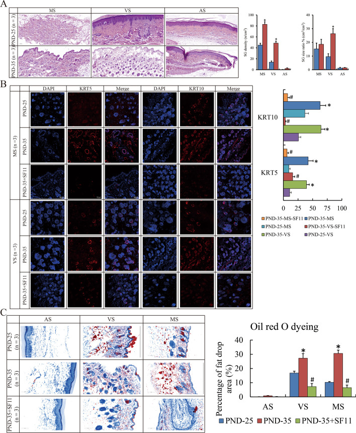
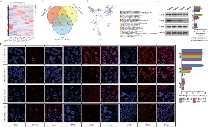
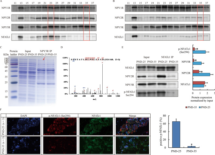
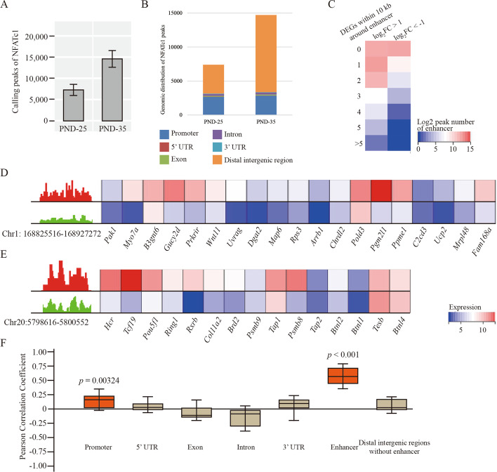
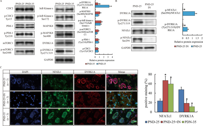
Methods: Herein, the rat volar and mammary tissues with abundant SGs from female SD rats with (post-natal day (PND)-35) and without puberty onset (PND-25) were arrested, and conducted RNA sequencing. The protein complex of Neuropeptide Y receptor Y2 (NPY2R)/NPY5R/Nuclear factor of activated T cells 1 (NFATc1) was performed by immunoprecipitation, mass spectrum and gel filtration. Genome-wide occupancy of NFATc1 was measured by chromatin immunoprecipitation sequencing. Target proteins' expression and localization was detected by western blot and immunofluorescence.

Results: NPY2R gene was significantly up-regulated in volar and mammary SGs of PND-25. A special protein complex of NPY2R/NPY5R/NFATc1 in PND-25. NFATc1 was dephosphorylated and activated, then localized into nucleus to exert as a transcription factor in volar SGs of PND-35. NFATc1 was especially binding at enhancer regions to facilitate the distal SG and sebum related genes' transcription. Dual specificity tyrosine phosphorylation regulated kinase 1A (DYRK1A) contributed to NFATc1 phosphorylation in PND-25, and inactivated of DYRK1A resulted in NFATc1 dephosphorylation and nuclear localization in PND-35.

Conclusions: Our findings unmask the new role of NPY2R/NFATc1/DYRK1A in pubertal SG, and are of benefit to advanced understanding the molecular mechanism of SGs' function after puberty, and provide some theoretical basis for the treatment of acne vulgaris from the perspective of hormone regulation.

Keywords: DYRK1A; NFATc1; NPY2R; NPY5R; Puberty onset; Sebaceous glands.


Conflict of interest statement

The authors declare that they have no competing interests.

Figures

Fig. 1
Fig. 1
Abundant SG distributed in pubertal skin. A H&E staining show the SG count and size in MS, VS and AS of PND-25 and PND-35 rat (magnification ×100). B IF show the expression of KRT5 and KRT10 in MS and VS of PND-25, PND-35 and PND-35 with SF-11 (magnification ×200). C Oil red O staining show the lipid droplet content in MS, VS and AS of PND-25, PND-35 and PND-35 with SF-11 (magnification ×200). The SG specimens from three individuals were used for immunofluorescence. Ten visual fields were randomly selected to count the positive staining, and calculate the proportion and statistical significance using Student’s t-test. “*” and “#” represent p-value less than 0.05 compared to the prior group. H&E: Hematoxylin and eosin staining; SG: sebaceous gland; PND: post-natal day; MS: mammary skin; VS: volar skin; AS: abdominal skin; IF: immunofluorescence; IP: immunoprecipitation; SF-11: NPY2R antagonist; DYRK1-IN-1/IN: DYRK1A inhibitor; DEGs: differential expressed genes; NFATc1: nuclear factor of activated T cells 1
Fig. 2
Fig. 2
Down-regulated NPY2R in pubertal skin. A Heatmap shows the DEGs of MS, VS an AS compared between PND-25 and PND-35. B Venn diagram shows the interested DEGs special for SG compared between PND-25 and PND-35. C Metascape analysis shows the GO and KEGG terms involved in interested DEGs for SG. D Western blot show the expression of NPY receptors in VS and MS of PND-25 and PND-35. E IF show the expression of NPY receptors in VS and MS of PND-25 and PND-35 (magnification with ×200). The SG specimens from two individuals were used for western blot and RNA sequencing. Statistical analysis and abbreviated description can be referred to Fig. 1 legend
Fig. 3
Fig. 3
Special protein complex of NPY2R/NPY5R/NFATc1 in SG before puberty onset. A, B Gel filtration assay show the distribution of NPY receptors and NFATc1 in VS of PND-25 (A) and PND-35 (B). Red frames highlighted the fraction of potential target protein peak. C Coomassie brilliant blue staining shows the proteome interacted with NPY5R in VS of PND-25 and PND-35. Red arrow highlights a specific band at 140 kDa only appears in PND-25. D Original secondary mass spectrum shows the most confident protein (NFATc1) identified by mass spectrum. E Western blot confirm the interaction between NFATc1 and NPY2R/NPY5R via phosphorylated form. F IF show the localization of NFATc1 and p-NFATc1 at Ser294 in VS of PND-25 and PND-35 (magnification ×200). The SG specimens from two individuals were used for gel filtration, immunoprecipitation and western blot. The SG specimens from three individuals were used for immunofluorescence. Statistical analysis and abbreviated description can be referred to Fig. 1 legend
Fig. 4
Fig. 4
Genome-wide occupancy of NFATc1 at enhancers in VS. A Overall calling peaks of NFATc1 in VS of PND-25 and PND-35 with two biological repetitions. B The genomic distribution of NFATc1 peaks. C Heatmap shows the count of DEGs (left: up-regulated, right: down-regulated DEGs) within 10 kb around differentially NFATc1 binding enhancers compared between PND-25 and PND-35. D, E IGV and heatmap show the two examples of NFATc1 peak at Chr1: 168825516–168927272 (D) as well as Chr20:5798616–5800552 (E) and the followed the transcription of gene within 10 kb distance in PND-25 and PND-35. F Pearson correlation analysis shows the coefficient of correlation between differential NFATc1 binding ability at each regions and target gene transcription. Red boxes indicate the coefficient with p-value less than 0.05 while brown ones indicate p-value without statistical significance. The SG specimens from two individuals were used for ChIP sequencing
Fig. 5
Fig. 5
The activity of NFATc1 induced by DYRK1A in early puberty. A Western blot show the expression of NFATc1 related phosphokinases and their phosphorylation in VS of PND-25 and PND-35. B Western blot show the phosphorylation of NFATc1 affected by DYRK1A inhibitor. C IF show the localization of NFATc1 and phosphorylated NFATc1 in VS of PND-25, PND25 with DYRK1A inhibitor and PND-35 (magnification ×200). The SG specimens from two individuals were used for western blot. The SG specimens from three individuals were used for immunofluorescence

Similar articles

Cited by

References

    1. Wang T, Zhou Z, Luo E, Zhong J, Zhao D, Dong H, Yao B. Comprehensive RNA sequencing in primary murine keratinocytes and fibroblasts identifies novel biomarkers and provides potential therapeutic targets for skin-related diseases. Cell Mol Biol Lett. 2021;26:42. doi: 10.1186/s11658-021-00285-6. - DOI - PMC - PubMed
    1. Zouboulis CC, Picardo M, Ju Q, Kurokawa I, Torocsik D, Biro T, Schneider MR. Beyond acne: current aspects of sebaceous gland biology and function. Rev Endocr Metab Disord. 2016;17:319–334. doi: 10.1007/s11154-016-9389-5. - DOI - PubMed
    1. Mollebjerg A, Palmen LG, Gori K, Meyer RL. The bacterial life cycle in textiles is governed by fiber hydrophobicity. Microbiol Spectr. 2021;9:e0118521. doi: 10.1128/Spectrum.01185-21. - DOI - PMC - PubMed
    1. Kippenberger S, Havlicek J, Bernd A, Thaci D, Kaufmann R, Meissner M. 'Nosing Around' the human skin: what information is concealed in skin odour? Exp Dermatol. 2012;21:655–659. doi: 10.1111/j.1600-0625.2012.01545.x. - DOI - PubMed
    1. Ludovici M, Kozul N, Materazzi S, Risoluti R, Picardo M, Camera E. Influence of the sebaceous gland density on the stratum corneum lipidome. Sci Rep. 2018;8:11500. doi: 10.1038/s41598-018-29742-7. - DOI - PMC - PubMed

Substances