

Primary pulmonary amoebiasis mimicking lung tumour in immunocompromised patient: A case report
- PMID: 37501684
- PMCID: PMC10368964
- DOI: 10.1002/rcr2.1199
Primary pulmonary amoebiasis mimicking lung tumour in immunocompromised patient: A case report
Abstract
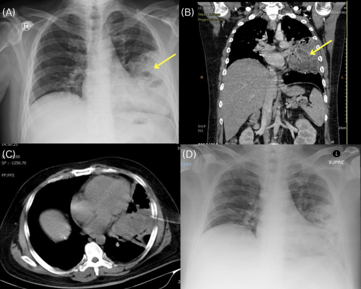
Amoebiasis is the most common protozoan disease caused by Entamoeba histolytica. The second most frequent extraintestinal infection, behind amoebic liver abscess, is pulmonary amoebiasis. We present the case of an immunocompromised 40-year-old man. He complained of cough for 1 month, shortness of breath, and fever. Chest x-ray demonstrated left paracardial consolidation, possibly pneumonia or a mass. Chest CT scans with contrast revealed the presence of an abscess-mimicking tumour. CT-guided TTB and histology examinations indicated the presence of trophozoites of E. histolytica. This patient was diagnosed with pulmonary amoebiasis. Diagnostic criteria for pulmonary amoebiasis include clinical manifestations, radiography, and microscopic examination. There was an improvement in clinical response after a 10-day course of antibiotics. Amoebiasis of the lungs is treatable with medicines and drainage when necessary. Early diagnosis and treatment are imperative to decrease mortality and morbidity.
Keywords: Amoebiasis; immunocompromised; parasitic; protozoan; pulmonary amoebiasis.
© 2023 The Authors. Respirology Case Reports published by John Wiley & Sons Australia, Ltd on behalf of The Asian Pacific Society of Respirology.
Conflict of interest statement
None declared.
Figures

References
-
- Pakrasi R, Mondal A, Kanungo C$A, Chakraborty M. Ruptured amoebic liver abscess into the left lung‐a case report [Internet]. Asian J Case Rep Surg. 2022;5(1):25–31. https://www.sdiarticle5.com/review-history/82048
-
- Dewi K, Suci Y, Dewi I, Iswanto I. Pulmonary amebiasis complicated with massive left empyema and respiratory failure: a case report. Sanamed [Internet]. 2020;15(1):45–49. https://scindeks.ceon.rs/Article.aspx?artid=1452-662X2001045D
-
- Aissa A, Hachicha M, Daadoucha A, Ben OI, Aissa S, Barhoumi H, et al. Pleuropulmonary amoebiasis: know to think about. J Pulm Respir Med [Internet]. 2017;7(3):409. https://www.omicsonline.org/open-access/pleuropulmonary-amoebiasis-know-...
-
- Samuel A, Binoy U, Ali A, Mohan N, Madhu K, Nair R, et al. Primary pulmonary amoebiasis. Pulmon the journal of respiratory sciences [Internet]. 2015;17:122–124. [Accessed October 20, 2022]. https://apccm.in/wp‐content/uploads/2017/02/PULMON‐SEP‐DEC‐15.pdf
Publication types
LinkOut - more resources
Full Text Sources
