Review
. 2023 Jun 24;1(4):356-371.
doi: 10.1021/cbmi.3c00036. eCollection 2023 Jul 24.

Application of Super-resolution SPEED Microscopy in the Study of Cellular Dynamics

Affiliations
Review

Application of Super-resolution SPEED Microscopy in the Study of Cellular Dynamics

Wenlan Yu et al. Chem Biomed Imaging. .

Abstract

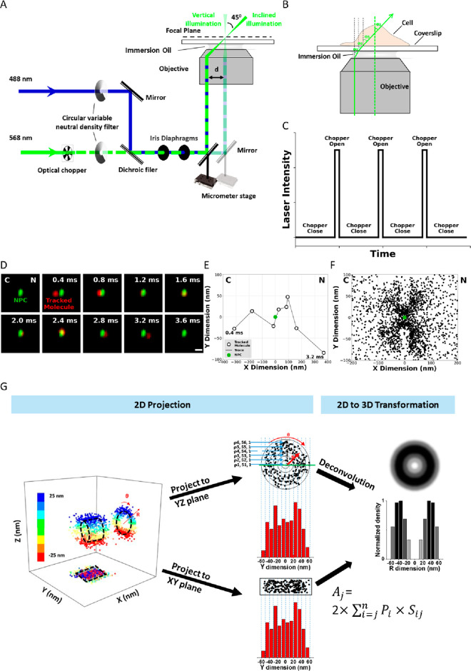
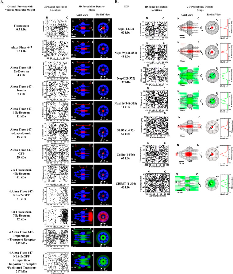
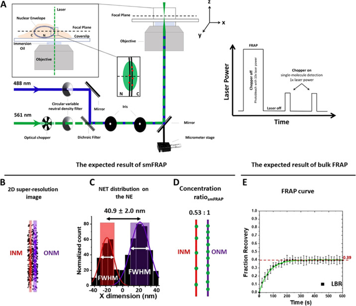
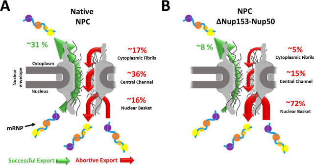
Super-resolution imaging techniques have broken the diffraction-limited resolution of light microscopy. However, acquiring three-dimensional (3D) super-resolution information about structures and dynamic processes in live cells at high speed remains challenging. Recently, the development of high-speed single-point edge-excitation subdiffraction (SPEED) microscopy, along with its 2D-to-3D transformation algorithm, provides a practical and effective approach to achieving 3D subdiffraction-limit information in subcellular structures and organelles with rotational symmetry. One of the major benefits of SPEED microscopy is that it does not rely on complex optical components and can be implemented on a standard, inverted epifluorescence microscope, simplifying the process of sample preparation and the expertise requirement. SPEED microscopy is specifically designed to obtain 2D spatial locations of individual immobile or moving fluorescent molecules inside submicrometer biological channels or cavities at high spatiotemporal resolution. The collected data are then subjected to postlocalization 2D-to-3D transformation to obtain 3D super-resolution structural and dynamic information. In recent years, SPEED microscopy has provided significant insights into nucleocytoplasmic transport across the nuclear pore complex (NPC) and cytoplasm-cilium trafficking through the ciliary transition zone. This Review focuses on the applications of SPEED microscopy in studying the structure and function of nuclear pores.


Conflict of interest statement

The authors declare no competing financial interest.

Figures

Figure 1
Figure 1
Basic setup of SPEED microscopy, working principles, and demonstration of single particle tracking, 2D to 3D transformation. (A) Schematic of the SPEED microscopy microscope setup for both the inclined and the vertical illumination. A 488 nm (blue) and a 568 nm (green) laser are directed into the objective in vertical illumination or offset so that the lasers pass through the focal plane at an angle of θ3. (B) Illustration of a molecule traveling in three dimensions as it passes through an NPC. A 2D projection of the same pathway is depicted in the lower panel. (C) Illustration of the effect of the optical chopper on laser intensity. The chopper is calibrated to be open for 1/10 of the frames captured. (D) Series of 2D locations of mRNA-mCherry (red spots) were captured by SPEED microscopy as they transported through the NPC (green spot). Numbers denote time in milliseconds. (E) Representative trajectories for successful nuclear transport of tracking single particle. (F) Experimentally determined 2D spatial locations of single molecule tracking in the NPC. (G) 3D information derived from 2D single-molecule data using a 2D-to-3D transformation. Adapted with permission from figures previously published by Nature Protocols.
Figure 2
Figure 2
Two-dimensional super-resolution spatial distributions and 3D transport routes of various (A) cytosol proteins and (B) IDPs with different molecular weights. Adapted with permission from figures previously published by PNAS and Protein Science.
Figure 3
Figure 3
Optical pathway for smFRAP as well as examples of expected data. (A) Schematic of the excitation laser used in smFRAP. A 561 nm laser (green) is directed into the objective. The inset depicts the laser passing through the nuclear envelope while the focal plane is at the nuclear equator, thereby illuminating NETs on both the inner and outer nuclear membrane. Inset illustrates laser power during the different phases of smFRAP. Fluorophores are initially photobleached using high laser power. The chopper is then engaged, allowing fluorescently tagged NETs to diffuse into the detection area. (B) Two-dimension (2D) super-resolution image of LBR on the NE, in which the INM shown in red and the ONM shown in purple. C) Two-peak Gaussian fittings of the points collected from the NE showing the distribution of LBR along the NE. The shaded regions represent the width of the INM and ONM as determined by the full width at half-maximum (fwhm) as determined by the fitting. (D) Approximate concentration ratios of LBR distribution along the INM (red) and ONM (purple). (E) FRAP curve for LBR. The mobile fraction of whole NE is ∼0.39. INM: inner nuclear membrane; ONM: inner nuclear membrane. Adapted with permission from figures previously published by Cell Press.
Figure 4
Figure 4
Nuclear export of mRNP through native (A) and aberrant NPCs (B). Both illustrations show an mRNP particle successfully exporting through the NPC (green arrow) and abortively exporting from the NPC (red arrow). The nuclear basket, central channel, or cytoplasmic fibril subregions separate the abortive nuclear export. The numerical values for the successful and abortive nuclear export indicate the transport efficiency from the 2021 Li et al. study.

References

    1. Singh B. K.; Nagar H.; Roichman Y.; Arie A. Particle manipulation beyond the diffraction limit using structured super-oscillating light beams. Light: Science & Applications 2017, 6 (9), e17050–e17050. 10.1038/lsa.2017.50. - DOI - PMC - PubMed
    1. Shakouri A.; Ziabari A.; Kendig D.; Bahk J. H.; Xuan Y.; Ye P. D.; Yazawa K.; Shakouri A.. Stable thermoreflectance thermal imaging microscopy with piezoelectric position control. In 2016 32nd Thermal Measurement, Modeling & Management Symposium (SEMI-THERM), 14–17 March 2016, 2016; pp 128–132. DOI:10.1109/SEMI-THERM.2016.7458456 - DOI
    1. Lelek M.; Gyparaki M. T.; Beliu G.; Schueder F.; Griffié J.; Manley S.; Jungmann R.; Sauer M.; Lakadamyali M.; Zimmer C. Single-molecule localization microscopy. Nature Reviews Methods Primers 2021, 1 (1), 1–27. 10.1038/s43586-021-00038-x. - DOI - PMC - PubMed
    1. von Diezmann L.; Shechtman Y.; Moerner W. Three-dimensional localization of single molecules for super-resolution imaging and single-particle tracking. Chem. Rev. 2017, 117 (11), 7244–7275. 10.1021/acs.chemrev.6b00629. - DOI - PMC - PubMed
    1. Gardill A.; Kemeny I.; Li Y.; Zahedian M.; Cambria M. C.; Xu X.; Lordi V.; Gali Á.; Maze J. R.; Choy J. T.; et al. Super-resolution airy disk microscopy of individual color centers in diamond. ACS Photonics 2022, 9 (12), 3848–3854. 10.1021/acsphotonics.2c00713. - DOI