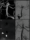
Preservation of Branching Vessel Using Super Compliant Double-Lumen Balloon Microcatheter: Bulging Neck Plasty Technique and Other Options
- PMID: 37501908
- PMCID: PMC10370972
- DOI: 10.5797/jnet.oa.2020-0152
Preservation of Branching Vessel Using Super Compliant Double-Lumen Balloon Microcatheter: Bulging Neck Plasty Technique and Other Options
Abstract
Objective: There are several methods to treat wide-neck aneurysms. We survey the cases that were treated using a super-compliant double-lumen balloon microcatheter (Super-Masamune) for preservation of the branching vessel originating proximal to the aneurysm, especially in the bulging neck plasty (BNP) technique.
Methods: We assessed 10 cases in which branching vessel preservation was performed using Super-Masamune. The cases were categorized into three groups: (1) ordinary neck plasty (ONP): balloon microcatheter was navigated to the branch that should be preserved; (2) BNP: another branch was preserved by inflating balloon bulging without cannulation; (3) protection during parent artery occlusion (PPO): the balloon microcatheter was navigated to the vessel to be occluded. The balloon preserves a branch originating near the aneurysm without cannulating to the branch.
Results: The aneurysm locations were as follows: internal carotid artery (ICA), three cases; anterior communicating artery (AcomA), one case; basilar artery (BA), three cases; and vertebral artery (VA), three cases. Four cases were ruptured aneurysms, while six cases were unruptured or ruptured in chronic stage. The ONP, BNP, and PPO groups contained two, five, and three cases, respectively. Embolization resulted in complete obliteration in six cases, neck remnant in two cases and body filling in two cases. No rupture/rerupture was noted in this series. One case showed an intraoperative rupture.
Conclusion: Super-Masamune is useful for neck plasty, especially BNP, in wide-neck aneurysms. Super-Masamune is also useful for parent artery occlusion when an important branch originates proximal to the aneurysm.
Keywords: balloon assist; embolization; neck plasty; parent artery occlusion.
©2021 The Japanese Society for Neuroendovascular Therapy.
Conflict of interest statement
We declare no conflicts of interest.
Figures


Similar articles
-
Intraaneurysmal Neck Plasty: Efficacy of a Super Compliant Double-Lumen Balloon Microcatheter.J Neuroendovasc Ther. 2021;15(5):301-309. doi: 10.5797/jnet.oa.2020-0148. Epub 2020 Dec 29. J Neuroendovasc Ther. 2021. PMID: 37501900 Free PMC article.
-
Modified balloon assisted coil embolization for the treatment of intracranial and cervical arterial aneurysms using coaxial dual lumen balloon microcatheters: initial experience.J Neurointerv Surg. 2014 Nov;6(9):704-7. doi: 10.1136/neurintsurg-2013-010936. Epub 2013 Oct 23. J Neurointerv Surg. 2014. PMID: 24153339
-
Balloon-in-stent technique for the constructive endovascular treatment of "ultra-wide necked" circumferential aneurysms.Neurosurgery. 2005 Dec;57(6):1218-27; discussion 1218-27. doi: 10.1227/01.neu.0000186036.35823.10. Neurosurgery. 2005. PMID: 16331170
-
Stent, balloon-assisted coiling and double microcatheter for treating wide-neck aneurysms in anterior cerebral circulation.Neurol Res. 2013 Dec;35(10):1002-8. doi: 10.1179/1743132813Y.0000000234. Epub 2013 Jun 20. Neurol Res. 2013. PMID: 23816570
-
[Coil Embolization for Very Small Intracranial Aneurysms with Diameter Less than 3mm:A Case Series of 14 Patients and Literature Review].No Shinkei Geka. 2018 Apr;46(4):303-312. doi: 10.11477/mf.1436203721. No Shinkei Geka. 2018. PMID: 29686163 Review. Japanese.
Cited by
-
Endovascular Treatment of Wide-Neck Bifurcation Aneurysm: Recent Trends in Coil Embolization with Adjunctive Technique.J Neuroendovasc Ther. 2024;18(3):75-83. doi: 10.5797/jnet.ra.2023-0072. Epub 2024 Jan 13. J Neuroendovasc Ther. 2024. PMID: 38559450 Free PMC article. Review.
-
Endovascular Treatments for Aneurysms Involving a Major Branch.J Neuroendovasc Ther. 2024;18(3):84-91. doi: 10.5797/jnet.ra.2023-0090. Epub 2024 Feb 22. J Neuroendovasc Ther. 2024. PMID: 38559454 Free PMC article. Review.
References
-
- Guglielmi G, Viñuela F, Dion J, et al. : Electrothrombosis of saccular aneurysms via endovascular approach. Part 2: Preliminary clinical experience. J Neurosurg 1991; 75: 8–14. - PubMed
-
- Ezura M, Kimura N, Uenohara H: Super-Masamune: developing process and animal study. JNET J Neuroendovasc Ther 2015; 9: 187–191. (in Japanese)
-
- Ezura M, Kimura N, Uenohara H: Super-Masamune: initial clinical experience. JNET J Neuroendovasc Ther 2015; 9: 192–196. (in Japanese)
-
- Takahashi A, Ezura M, Yoshimoto T: Broad neck basilar tip aneurysm treated by neck plastic intra-aneurysmal GDC embolisation with protective balloon. Interv Neuroradiol 1997; 3: 167–170. - PubMed
LinkOut - more resources
Full Text Sources
Miscellaneous

