. 2023 Jul 19:2023:6623329.
doi: 10.1155/2023/6623329. eCollection 2023.

Vitronectin Destroyed Intestinal Epithelial Cell Differentiation through Activation of PDE4-Mediated Ferroptosis in Inflammatory Bowel Disease

Affiliations

Vitronectin Destroyed Intestinal Epithelial Cell Differentiation through Activation of PDE4-Mediated Ferroptosis in Inflammatory Bowel Disease

Wenxu Pan et al. Mediators Inflamm. .

Abstract

Objective: Vitronectin (VTN) has been reported to trigger cell pyroptosis to aggravate inflammation in our previous study. However, the function of VTN in inflammatory bowel disease (IBD) remains to be addressed.

Methods: Real-time PCR and western blotting were performed to analyze VTN-regulated intestinal epithelial cell (IEC) differentiation through ferroptosis, and immunofluorescence (IF), luciferase, and chromatin immunoprecipitation were used to identify whether VTN-modulated ferroptosis is dependent on phosphodiesterase 4 (PDE4)/protein kinase A (PKA)/cyclic adenosine monophosphate-response element-binding protein (CREB) cascade pathway. In vivo experiment in mice and a pilot study in patients with IBD were used to confirm inhibition of PDE4-alleviated IECs ferroptosis, leading to cell differentiation during mucosal healing.

Results: Herein, we found that caudal-related homeobox transcription factor 2-mediated IECs differentiation was impaired in response to VTN, which was attributed to enhanced ferroptosis characterized by decreased glutathione peroxidase 4 (GPX4) and solute carrier family 7 member 11 expression. Inhibition of ferroptosis in IECs rescued the inhibitory effect of VTN on cell differentiation. Further analysis showed that VTN triggered phosphorylation of PDE4, leading to inhibit PKA/CREB activation and CREB nuclear translocation, which further reduced GPX4 transactivation. Endogenous PKA interacted with CREB, and this interaction was destroyed in response to VTN stimulation. What is more, overexpression of CREB in CaCO2 cells overcame the promotion of VTN on ferroptosis. Most importantly, inhibition of PDE4 by roflumilast or dipyridamole could alleviate dextran sulfate sodium-induced colitis in mice and in a pilot clinical study confirmed by IF.

Conclusions: These findings demonstrated that highly expressed VTN disrupted IECs differentiation through PDE4-mediated ferroptosis in IBD, suggesting targeting PDE4 could be a promising therapeutic strategy for patients with IBD.


Conflict of interest statement

The authors declare that they have no conflicts of interest.

Figures

Figure 1
Figure 1
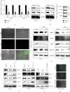
VTN suppressed intestinal epithelial cell differentiation. (a) Immunofluorescence of VTN expression level in indicated intestinal tissue from clinical sample and colitis model. (b) Real-time PCR and (c) Western blotting and quantified analysis were performed to detect the effect of VTN on CDX2 and MUC2 expression. Data presented as the mean ± s.e.m. of three independent experiments and were analyzed by t test,  ∗∗∗∗p < 0.0001. (d) Intestinal organoid assay was used to explore the effect of VTN on intestinal organoid generation. (e) The immunofluorescence was performed to analyze CDX2 and MUC2 expression in intestinal organoid in response to VTN stimulation described as (d).
Figure 2
Figure 2
VTN promoted ferroptosis to suppress cell differentiation. (a) Intestinal epithelial cells were digested and reseeded in a 6-well plate overnight; after 12-serum-free treatment, the cells were treated with or without VTN (5 μg/ml) for another 48 hr, real-time PCR were performed to analyze GPX4 and SLC7A11 expression at mRNA level, data presented as the mean ± s.e.m. of three independent experiments and were analyzed by t test,  ∗∗∗∗p < 0.0001. (b) CaCO2 and HT-29 cells were treated and described as (a). The total protein was collected and subjected by western blotting to detect GPX4 and SLC7A11 expression at protein level; the band intensity was measured and quantified by t test, data presented as the mean ± s.e.m. of three independent experiments and were analyzed by t test,  ∗∗∗p < 0.001,  ∗∗∗∗p < 0.0001. (c) CaCO2 cells were treated described as (a), and the ROS level was detected according to the manufacturer's instruction. (d) After starvation, CaCO2 and HT-29 cells were treated as indicated for 48 hr, and the total protein level was collected; WB was employed to detected activation of ferroptosis by RSL3 on intestinal epithelial cell differentiation. (e) After serum starvation for 24 hr, CaCO2 and HT-29 cells were treated with or without VTN, followed by ferroptosis inhibitor ferrostatin-1 (Fer) (10 μM) stimulation for further 48 hr. The total lysates were collected to analyze indicated protein levels. (f) 24 hr after transfection with GPX4 plasmid, CaCO2 cells were treated with or without VTN for another 24 hr to collect the total protein; WB was performed to detect indicated proteins. (g) CaCO2 cells were treated as (f), and ROS staining was used to analyze the ROS level.
Figure 3
Figure 3
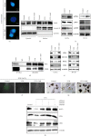
VTN regulated ferroptosis through CREB. (a) Internal control pGL4.17 and GPX4 promoter reporter plasmid together with pCDNA 3.0 or CREB plasmid were transfected into HT-29 cells, followed by treated with or without VTN for 48 hr, relative luciferase activities unit were measured and analyzed by two-way analysis of variance (ANOVA) and Dunnett's multiple comparison test,  ∗∗∗p < 0.001,  ∗∗∗∗p < 0.0001, n = 3, data presented as the mean ± s.e.m. (b) ChIP analysis of binding of CREB protein to GPX4 gene promoter in CaCO2 cells treated as indicated. Student's t-test, data presented as the mean ± s.e.m, relative luciferase activities unit were measured and analyzed by two-way analysis of variance (ANOVA) and Dunnett's multiple comparison test,  ∗∗∗p < 0.001,  ∗∗∗∗p < 0.0001, n = 3. (c) HT-29 cells were transfected with or without CREB for 24 hr, further incubation with VTN was performed for another 24 hr, the total lysate was collected and subjected from WB to examine indicated protein.
Figure 4
Figure 4
VTN modulated PDE4/PKA/CREB signaling. (a) Immunofluorescence was employed to display the CREB nuclear localization in CaCO2 cells treated with VTN or VTN combined with roflumilast (Rofl) for 1 hr. (b) CaCO2 cells were serum starved for 24 hr and then stimulated as indicated for an additional 1 hr. Nuclear and cytosolic CREB levels were determined by western blotting. GAPDH and lamin A/B were served as internal controls for the cytosolic and nuclear fractions, respectively. (c) After serum starved for 24 hr, CaCO2 and HT-29 cells were treated with or without VTN for 1 hr, the total proteins were collected and separated to detect indicated protein by western blotting. α-Tubulin was taken as internal control. (d) HT-29 cells were serum starved for 24 hr after 80% confluence, then stimulated as indicated for 1 hr. Immunoprecipitated (IP) was employed to analyze the interaction between PKA and CREB in response to VTN and VTN combined with Rofl. (e) After serum starved for 24 hr, the total cell was pretreated with Rofl for 1 hr; subsequently, followed by stimulation with or without VTN for further 48 hr, the total protein was collected to detect indicated protein. α-tubulin was served as internal control. (f) CaCO2 cells were treated with basic medium for 24 hr, followed by stimulation with VTN and VTN combined with Rofl for further 48 hr, the level of ROS was detected. (g) Intestinal organoids assay was performed to analyze the effect of VTN-Rofl on cell differentiation. (h) After transfection with indicated siRNA for 24 hr, HT-29 cells were starved for 12 hr and treated with or without VTN for another 24 hr; the total protein was collected to detect indicated proteins.
Figure 5
Figure 5
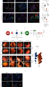
Inhibition of PDE4 alleviated colitis. In a DSS-induced acute colitis model, mice were treated with dipyridamole (10 mg/kg) daily for 1 month, and the body weight was recorded every 2 days (a) The statistical difference was performed by one-way ANOVA,  ∗∗∗∗p < 0.0001, control, n = 6; DSS + vehicle, n = 6; DSS + dipyridamole, n = 6, data presented as the mean ± s.e.m. (b) The representative image of colon length was captured and the statical analysis were performed by one-way ANOVA,  ∗∗∗∗p < 0.0001, data are represented as mean ± s.e.m.;  ∗∗∗∗p < 0.0001. (c) Immunofluorescence of indicated protein expression level was used to show the effect of DIP in DSS-induced colitis, quantitation was performed by Image J, and data presented as the mean ± s.e.m and analyzed by one-way ANOVA,  ∗∗∗∗p < 0.0001, MFI: mean fluorescence intensity. (d) Schematic view of IBD patients enrolled in a pilot clinical study received endoscopy assessment and biopsy collection. The information for the pilot study is provided in Table 1. (e) Analysis of clinical disease activity index (DAI) evaluation in five IBD children in indicated timepoint. Data are represented as mean ± s.e.m; p values were calculated by t test. n = 5,  ∗∗∗∗p < 0.0001. (f) The representative endoscopic images of before and after DIP administration in five children with IBD, and score for endoscopy was evaluated. (g) Immunofluorescence of indicated protein expression level was used to show the effect of DIP in patients with IBD, quantitation was performed by Image J, and data presented as the mean ± s.e.m and analyzed by paired t test,  ∗∗∗∗p < 0.0001, MFI: mean fluorescence intensity. (h) Schematic model of VTN-regulated intestinal epithelial cell differentiation through ferroptosis.

References

    1. Zhang S., Xu W., Wang H., et al. Inhibition of CREB-mediated ZO-1 and activation of NF-κB-induced IL-6 by colonic epithelial MCT4 destroys intestinal barrier function. Cell Proliferation . 2019;52(6) doi: 10.1111/cpr.12673.e12673 - DOI - PMC - PubMed
    1. Huang L., Tang X., Yang F. Y., et al. Shikonin contributes to intestinal epithelial cell differentiation through PKM2/NRF2-mediated polyol pathway. Pharmacological Research – Modern Chinese Medicine . 2021;1 doi: 10.1016/j.prmcm.2021.100004.100004 - DOI
    1. Sun X., Yang Q., Rogers C. J., Du M., Zhu M.-J. AMPK improves gut epithelial differentiation and barrier function via regulating Cdx2 expression. Cell Death & Differentiation . 2017;24:819–831. doi: 10.1038/cdd.2017.14. - DOI - PMC - PubMed
    1. Wang Q., Zhou Y., Rychahou P., et al. Ketogenesis contributes to intestinal cell differentiation. Cell Death & Differentiation . 2017;24:458–468. doi: 10.1038/cdd.2016.142. - DOI - PMC - PubMed
    1. Takayama K., Negoro R., Yamashita T., et al. Generation of human iPSC–derived intestinal epithelial cell monolayers by CDX2 Transduction. Cellular and Molecular Gastroenterology and Hepatology . 2019;8(3):513–526. doi: 10.1016/j.jcmgh.2019.06.004. - DOI - PMC - PubMed

Substances