Review
. 2020;14(12):583-592.
doi: 10.5797/jnet.ra.2020-0131. Epub 2020 Nov 25.

Direct Carotid Cavernous Fistulas

Affiliations
Review

Direct Carotid Cavernous Fistulas

Satomi Ide et al. J Neuroendovasc Ther. 2020.

Abstract

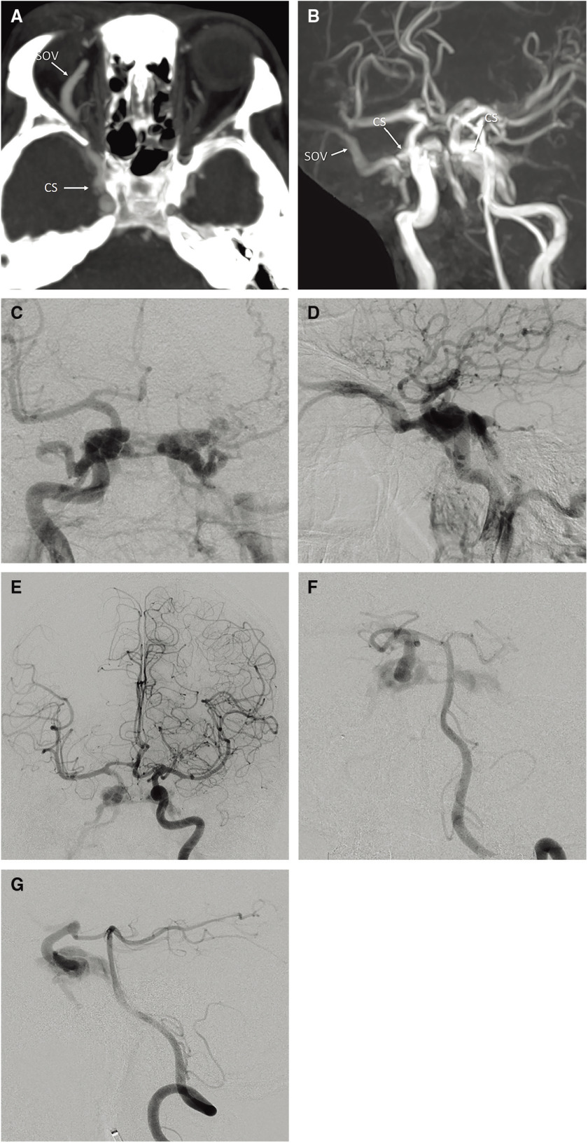
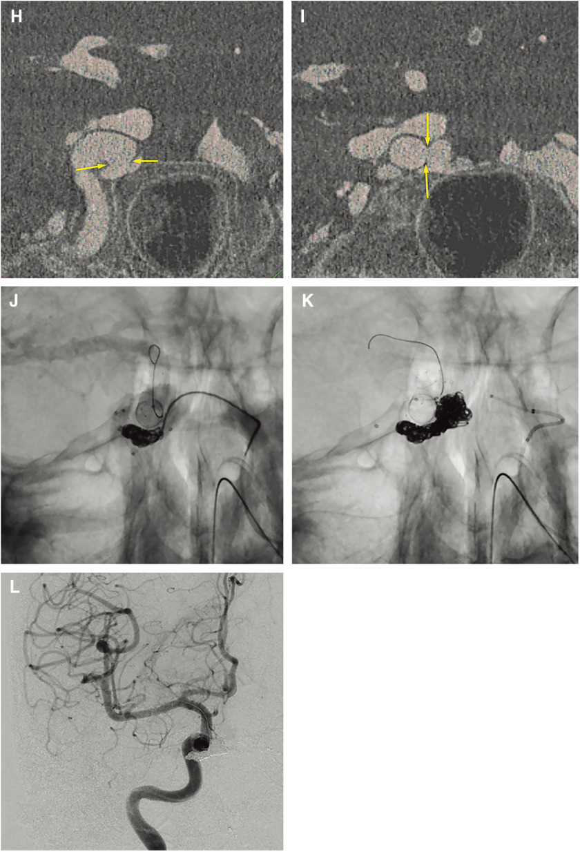
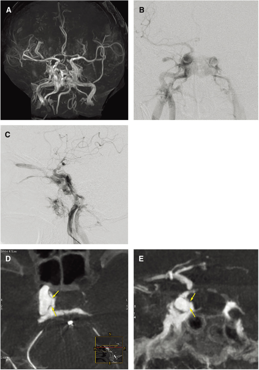
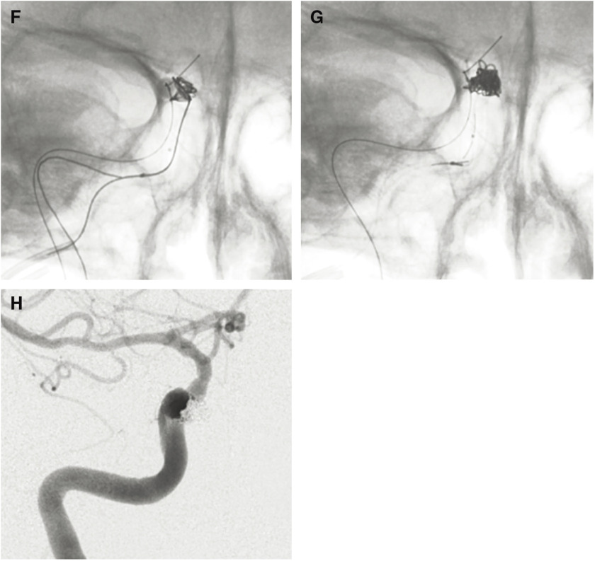
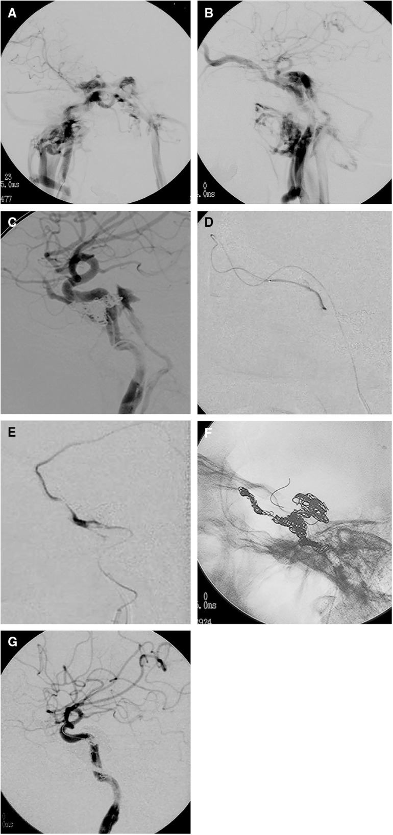
A direct carotid cavernous fistula (CCF) is an abnormal shunt between the internal carotid artery (ICA) and the cavernous sinus (CS). Traumatic CCF is the most common type, accounting for up to 75% of all CCFs. For the management of direct CCF, endovascular therapy has become the standard. For successful endovascular therapy, evaluation of the size and location of orifice of the CCF, venous drainage, and tolerance for ICA occlusion on cerebral angiography is necessary. Multi-planner reformatted images of 3D rotation angiography are useful to visualize the fistula and compartments of the CS precisely. Due to the limited commercial availability of detachable balloons, detachable coils have become a widely employed endovascular tool for the treatment of direct CCFs. The advantageous aspects of coil application are their easy retrievability and better control. In the case of large/multiple fistulas, adjunctive techniques, including balloon- and stent-assisted techniques, are often needed to occlude the CCF while preserving the ICA. To avoid cranial nerve palsy related to over-packing of the CS with detachable coils or a detachable balloon, selective embolization of the fistula portion is required. Use of liquid embolic materials and covered stents was recently reported as another adjunctive technique. In cases in which it is impossible to occlude the CCF while preserving the ICA, parent artery occlusion (PAO) is considered. The selection of additional/alternative techniques and devices depends on the anatomy and hemodynamics of each CCF, and the skill and experience of individual operators.

Keywords: coil embolization; detachable balloon; direct carotid cavernous fistula; transarterial embolization; transvenous embolization.


Conflict of interest statement

The authors declare that they have no conflict of interest.

Figures

Fig. 1
Fig. 1. A 43-year-old male presented with proptosis, chemosis, and diplopia after a traffic accident. (A) Axial partial MIP enhanced CT image showing dilatation of the right SOV and the right CS. (B) Right anterior oblique view on 3D-TOF MRA showing the dilated right SOV and the bilateral CSs. (C, D) Frontal (C) and lateral (D) views on right internal carotid angiography showing a direct high-flow shunt between the horizontal intracavernous segment of the right ICA and the CS. The direct CCF drained into the contralateral CS via the inter-CS, the bilateral SOVs, and the bilateral inferior petrosal sinuses. The left superficial middle cerebral vein and the superior petrosal sinus drained retrogradely into the bilateral superior ophthalmic vein and bilateral inferior petrosal sinus. On the left side, dangerous cerebral venous refluxes, including the superficial middle cerebral vein, uncal vein, and the petrosal vein, were observed. (E) Left internal carotid angiography with the Matas maneuver showing collateral flow to the right anterior and middle cerebral arteries via the anterior communicating artery and retrograde filling to the CCF. The right anterior and middle cerebral arteries are not sufficiently opaque compared with the contralateral counterparts due to arterial steal to the CCF. (F, G) Frontal (F) and lateral (G) views on left vertebral angiography with the Allcock maneuver showing collateral flow via the posterior communicating artery. The location and size of the fistula were more clearly visualized. 3D-TOF: 3D time of flight; CCF: carotid cavernous fistula; CS: cavernous sinus; ICA: internal carotid artery; MIP: maximum intensity projection; SOV: superior ophthalmic vein (H, I) Coronal reconstructed 3D rotational angiography images of the right ICA (H, I; from the dorsal to ventral side) clearly showing two fistulas (yellow arrows) located at the inner wall and inferior wall of the horizontal cavernous portion of the right ICA. The CCF was treated by selective coil embolization using balloon- and stent-assisted techniques. Via both transarterial and transvenous approaches, two microcatheters were transvenously introduced into a fistulous pouch located inferiorly to the ICA, and another microcatheter was introduced into the same pouch transarterially. Coils were first placed into the fistulous pouch under the balloon- assisted technique. (J) Fluoroscopic image from the frontal view during balloon-assisted coil embolization showing that coils were introduced via three microcatheters (one via the right IPS, another via the left IPS, and the other via the ICA) placed in the same fistulous pouch located inferiorly to the ICA. (K) Fluoroscopic image from the frontal view after embolization. As there were two fistulas and the inferior one was too large to sufficiently pack the coils densely, selective coil embolization was continued using the stent-assisted technique. (L) Right internal carotid angiography after embolization showing complete occlusion of the CCF while preserving the right ICA. CCF: carotid cavernous fistula; ICA: internal carotid artery; IPS: inferior petrosal sinus; (The color version is available online.)
Fig. 2
Fig. 2. A 91-year-old female with traumatic direct CCFs. (A) Coronal view on 3D-TOF MRA showing the high signal intensity of the bilateral CS and the right SOV. (B, C) Frontal (B) and lateral (C) views on right internal carotid angiography showing the high-flow CCF at the right cavernous portion. The direct CCF drained into the right CS to the contralateral CS, the bilateral inferior petrosal sinus, the bilateral pterygoid plexus, and the bilateral SOVs. (D, E) Axial (D) and coronal (E) reconstructed 3D angiography images of the right ICA clearly depicting the location of the fistula (yellow arrows) and fistulous venous pouch medially to the ICA. The direct CCF was treated by balloon-assisted coil embolization. A micro balloon catheter was placed crossing over the fistula in the cavernous portion of the right ICA and a microcatheter was advanced transarterially into the fistula via the orifice. Via the transvenous approach, two microcatheters were advanced into the same fistulous pouch via the right inferior petrosal sinus. 3D-TOF: 3D time of flight; CCF: carotid cavernous fistula; CS: cavernous sinus; ICA: internal carotid artery; SOV: superior ophthalmic vein; (The color version is available online.) (F) Fluoroscopic image showing three microcoils from three different microcatheters that were deployed into the fistula compartment under balloon inflation. (G) The fistulous pouch was packed with nine coils. (H) Right internal carotid angiography after selective embolization showing complete occlusion of the CCF. CCF: carotid cavernous fistula;
Fig. 3
Fig. 3. A case of traumatic CCF with delayed abducens nerve palsy that developed 5 years after embolization by CS packing with coils. (A, B) Frontal (A) and lateral (B) views on right internal carotid angiography showing a high-flow direct CCF draining into the right SOV, right superior petrosal sinus, contralateral CS via the inter-CS, bilateral IPS, and bilateral pterygoid plexus. A microcatheter was advanced into the CS via the fistula and the posterior compartment of the CS was embolized using coils. (C) Lateral view on right internal carotid angiography after transarterial embolization showing a small residual CCF draining into the right inferior petrosal sinus and the right SOV. Right abducens nerve palsy developed a few days after embolization. As the CCF remained on follow-up angiography a few weeks later, repeated embolization was performed via both transvenous and transarterial approaches. (D) Selective angiography of the CS via the catheter introduced via the right superficial temporal vein, angular vein, and the SOV. The anterior part of the CS and the SOV was embolized using coils. Then, the microcatheter was advanced via the right inferior petrosal sinus into the right superficial middle cerebral vein. (E) Selective angiography of the right superficial middle cerebral vein showing cortical venous reflux. The proximal portion of the superficial middle cerebral vein and the lateral compartment of the CS were embolized using coils. Then, a microcatheter was advanced from the ICA via the tear into the posterior compartment of the CS, and the compartment was embolized using coils under the balloon-assisted technique. (F) Lateral views on fluoroscopy at the end of the embolization procedures showing the coil mass in the entire CS. (G) Lateral view on right internal carotid angiography showing complete occlusion of the CCF. The patient’s symptoms completely resolved within 3 months after embolization. However, right abducens nerve palsy recurred 5 years after embolization. Repeated angiography and head MRI demonstrated no recurrence of the CCF or other abnormal findings. The recurrent abducens nerve palsy became a permanent deficit. 3D-TOF: 3D time of flight; CCF: carotid cavernous fistula; CS: cavernous sinus; ICA: internal carotid artery; IPS: inferior petrosal sinus; SOV: superior ophthalmic vein

References

    1. Lylyk P, Viñuela F, Campos J, et al. : Diagnosis and endovascular therapy of vascular lesions in the cavernous sinus. Acta Radiol Suppl 1986; 369: 584–585. - PubMed
    1. Fleishman JA, Garfinkel RA, Beck RW, et al. : Advances in the treatment of carotid cavernous fistulas. Int Ophthalmol Clin 1986; 26: 301–311. - PubMed
    1. Barrow DL, Spector RH, Braun IF, et al. : Classification and treatment of spontaneous carotid-cavernous sinus fistulas. J Neurosurg 1985; 62: 248–256. - PubMed
    1. Ringer AJ, Salud L, Tomsick TA: Carotid cavernous fistulas: anatomy, classification, and treatment. Neurosurg Clin N Am 2005; 16: 279–295. - PubMed
    1. Van Overmeire O, De Keukeleire K, Van Langenhove P, et al. : Carotid-cavernous fistula in ehlers-danlos syndrome by pure transvenous approach. Interv Neuroradiol 2006; 12: 45–51. - PMC - PubMed