

doi: 10.54844/prm.2022.0104.
Epub 2022 Nov 28.
Fetal membrane at the feto-maternal interface: An underappreciated and understudied intrauterine tissue
Affiliations
- PMID: 37502422
- PMCID: PMC10373051
- DOI: 10.54844/prm.2022.0104
Item in Clipboard
Fetal membrane at the feto-maternal interface: An underappreciated and understudied intrauterine tissue
Placenta Reprod Med.
.
No abstract available
Conflict of interest statement
Conflict of Interest Ramkumar Menon is an Editorial Board Member of the journal. The article was subject to the journal’s standard procedures, with peer review handled independently of this member and her research group.
Figures
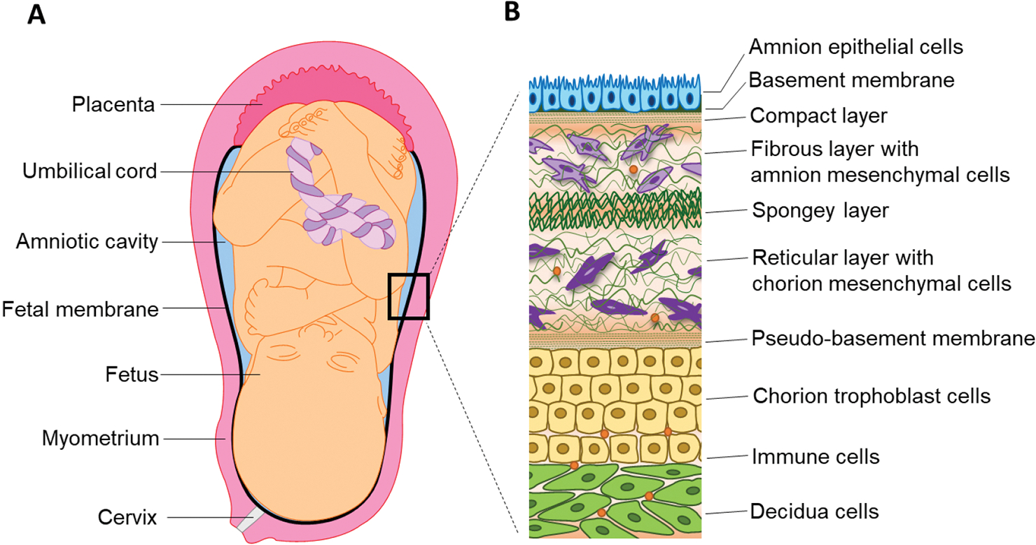
Intrauterine and fetal membrane anatomy. (A) Within the intrauterine cavity, there are a variety of maternal (i.e., myometrium and cervix) and fetal (i.e., placenta, umbilical cord, and an amniotic cavity containing amniotic fluid, and the fetal membranes) derived organs that surround the fetus and contribute to pregnancy maintenance. The fetal membranes (black) line the cavity and are derived from multiple fetal cellular and collagen layers to form the feto-maternal interface. (B) The amnion epithelial cells (blue) are connected to the basement membrane (dark green) and compact layer (green dashes) of the extracellular matrix (ECM) forming an amniotic fluid-tight barrier. Within the first layer of the ECM (i.e., the fibrous layer), amnion mesenchymal cells (light purple) migrate and interact with the collagen environment. Separating the fibrous and reticular layers of the ECM is the spongy layer that separates the amnion (blue) and chorion (yellow) portions of the fetal membranes. The reticular layer of the ECM contains chorion mesenchymal cells (dark purple) that is connected to the pseudo-basement membrane of the chorion. The multi-layer of chorion trophoblast cells (yellow) forms the second epithelial layer of the fetal membranes and is critical for immune homeostasis. The fetal chorion layer is directly connected to the maternal decidua layer (green) forming the feto-maternal interface of the membranes. Resident immune cells predominantly live in the decidua layer but can migrate into the chorion and amnion layers if stimulated.
Similar articles
-
Organic Anion Transporting Polypeptide 2B1 in Human Fetal Membranes: A Novel Gatekeeper for Drug Transport During Pregnancy?Front Pharmacol. 2021 Dec 20;12:771818. doi: 10.3389/fphar.2021.771818. eCollection 2021. Front Pharmacol. 2021. PMID: 34987396 Free PMC article.
-
Organ-On-Chip Technology: The Future of Feto-Maternal Interface Research?Front Physiol. 2020 Jun 30;11:715. doi: 10.3389/fphys.2020.00715. eCollection 2020. Front Physiol. 2020. PMID: 32695021 Free PMC article. Review.
-
Intermembrane distances at the feto-maternal interface in epitheliochorial placentation.Placenta. 2021 Jun;109:37-42. doi: 10.1016/j.placenta.2021.04.011. Epub 2021 Apr 24. Placenta. 2021. PMID: 33965813
-
[Feto-maternal transfusion following cordocentesis].Orv Hetil. 1998 Aug 30;139(35):2059-64. Orv Hetil. 1998. PMID: 9755624 Hungarian.
-
New insights into myeloid-derived suppressor cells and their roles in feto-maternal immune cross-talk.J Reprod Immunol. 2016 Feb;113:35-41. doi: 10.1016/j.jri.2015.11.001. Epub 2015 Nov 10. J Reprod Immunol. 2016. PMID: 26599285 Review.
Cited by
-
Escherichia coli induced matrix metalloproteinase-9 activity and type IV collagen degradation is regulated by progesterone in human maternal decidual.BMC Pregnancy Childbirth. 2024 Oct 4;24(1):645. doi: 10.1186/s12884-024-06847-8. BMC Pregnancy Childbirth. 2024. PMID: 39367340 Free PMC article.
-
Biological importance of human amniotic membrane in tissue engineering and regenerative medicine.Mater Today Bio. 2023 Sep 1;22:100790. doi: 10.1016/j.mtbio.2023.100790. eCollection 2023 Oct. Mater Today Bio. 2023. PMID: 37711653 Free PMC article. Review.
-
Determining Sex-Specific Gene Expression Differences in Human Chorion Trophoblast Cells.Int J Mol Sci. 2025 Mar 2;26(5):2239. doi: 10.3390/ijms26052239. Int J Mol Sci. 2025. PMID: 40076861 Free PMC article.
-
Analysis of the Protective Potential of the Amniotic Membrane in an In Vitro Experimental Model of Demyelination in Mouse Brain Organotypic Slices.ACS Omega. 2025 Jul 23;10(30):33162-33177. doi: 10.1021/acsomega.5c02999. eCollection 2025 Aug 5. ACS Omega. 2025. PMID: 40787362 Free PMC article.
References
-
- Menon R, Moore JJ. Fetal membranes, not a mere appendage of the placenta, but a critical part of the fetal-maternal interface controlling parturition. Obstet Gynecol Clin North Am 2020;47:147–162. - PubMed
Grants and funding
LinkOut - more resources
Full Text Sources