Case Reports
. 2022;16(4):225-231.
doi: 10.5797/jnet.cr.2021-0021. Epub 2021 Sep 3.

Non-Sinus-Type Laterocavernous Sinus Dural Arteriovenous Fistula Treated by Transarterial Venous Coil Embolization: A Case Report

Affiliations
Case Reports

Non-Sinus-Type Laterocavernous Sinus Dural Arteriovenous Fistula Treated by Transarterial Venous Coil Embolization: A Case Report

Satoshi Inoue et al. J Neuroendovasc Ther. 2022.

Abstract

Objective: Laterocavernous sinus dural arteriovenous fistulas (DAVFs) are rare and not always accessible transvenously due to their angioarchitecture. We report a case of non-sinus-type laterocavernous sinus DAVF treated by endovascular transarterial venous coil embolization.

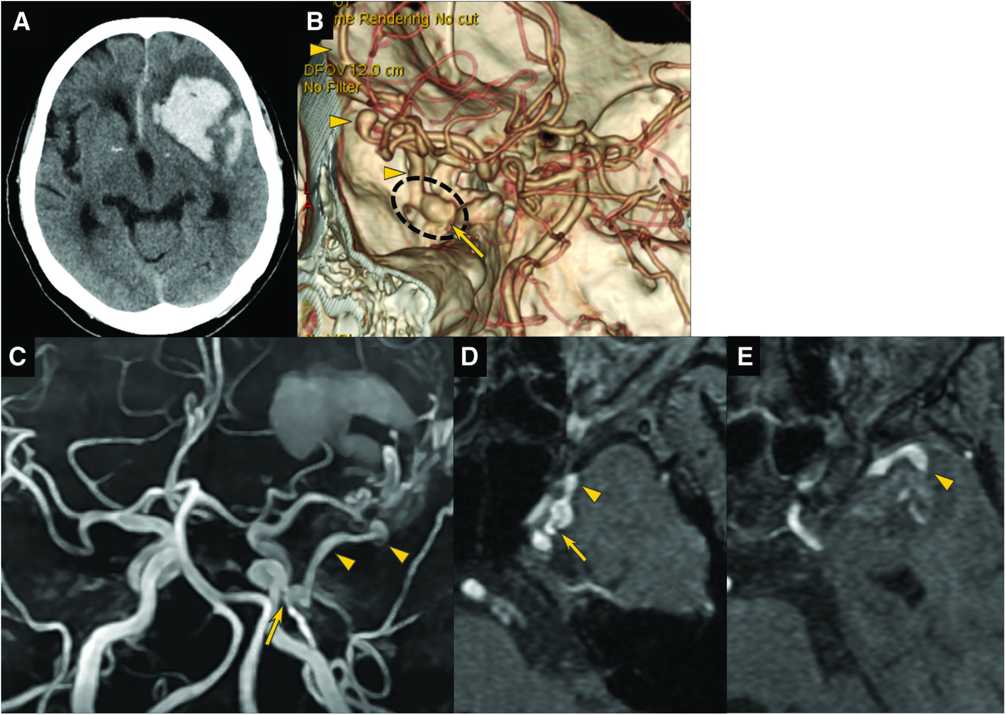
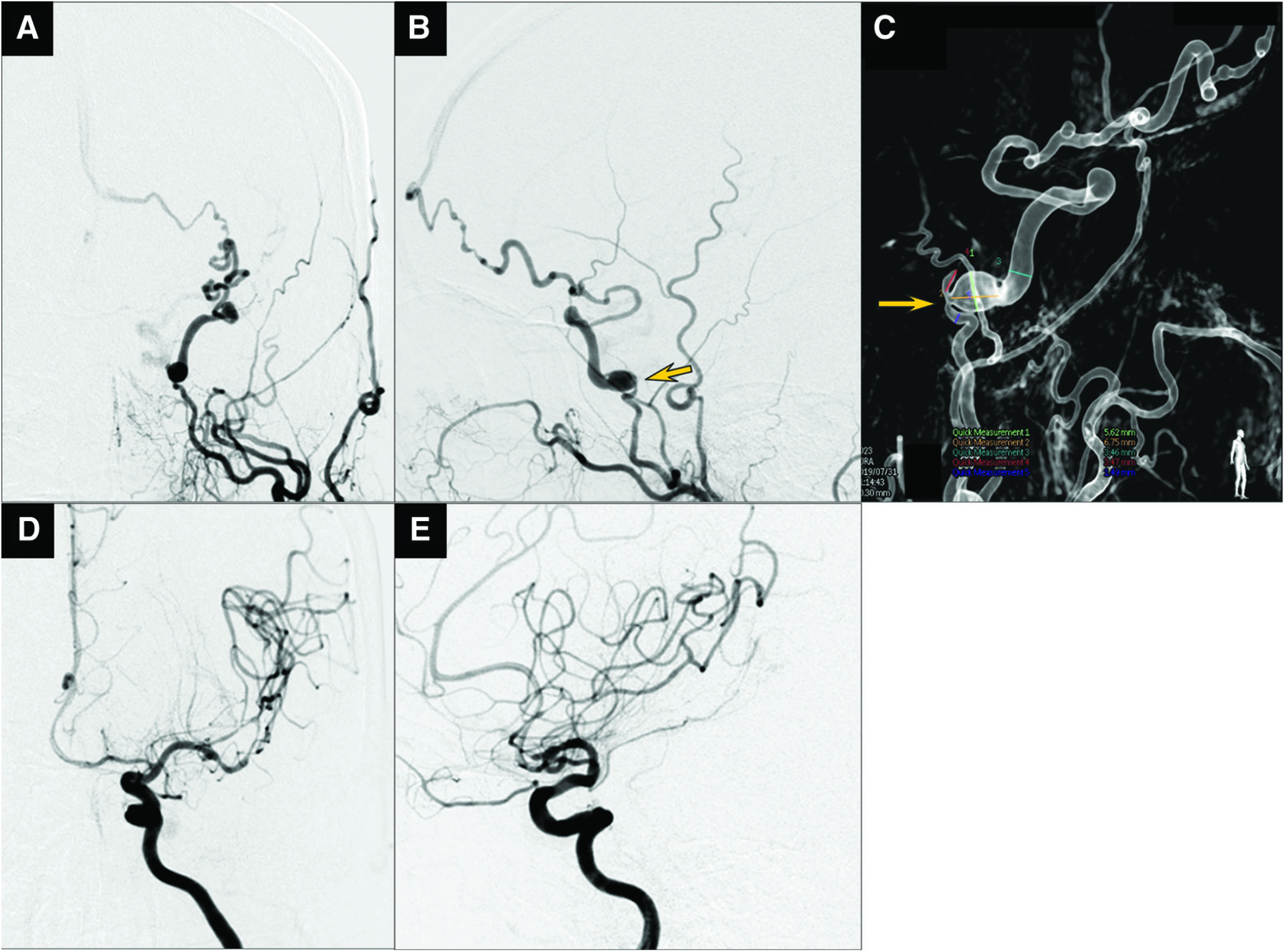
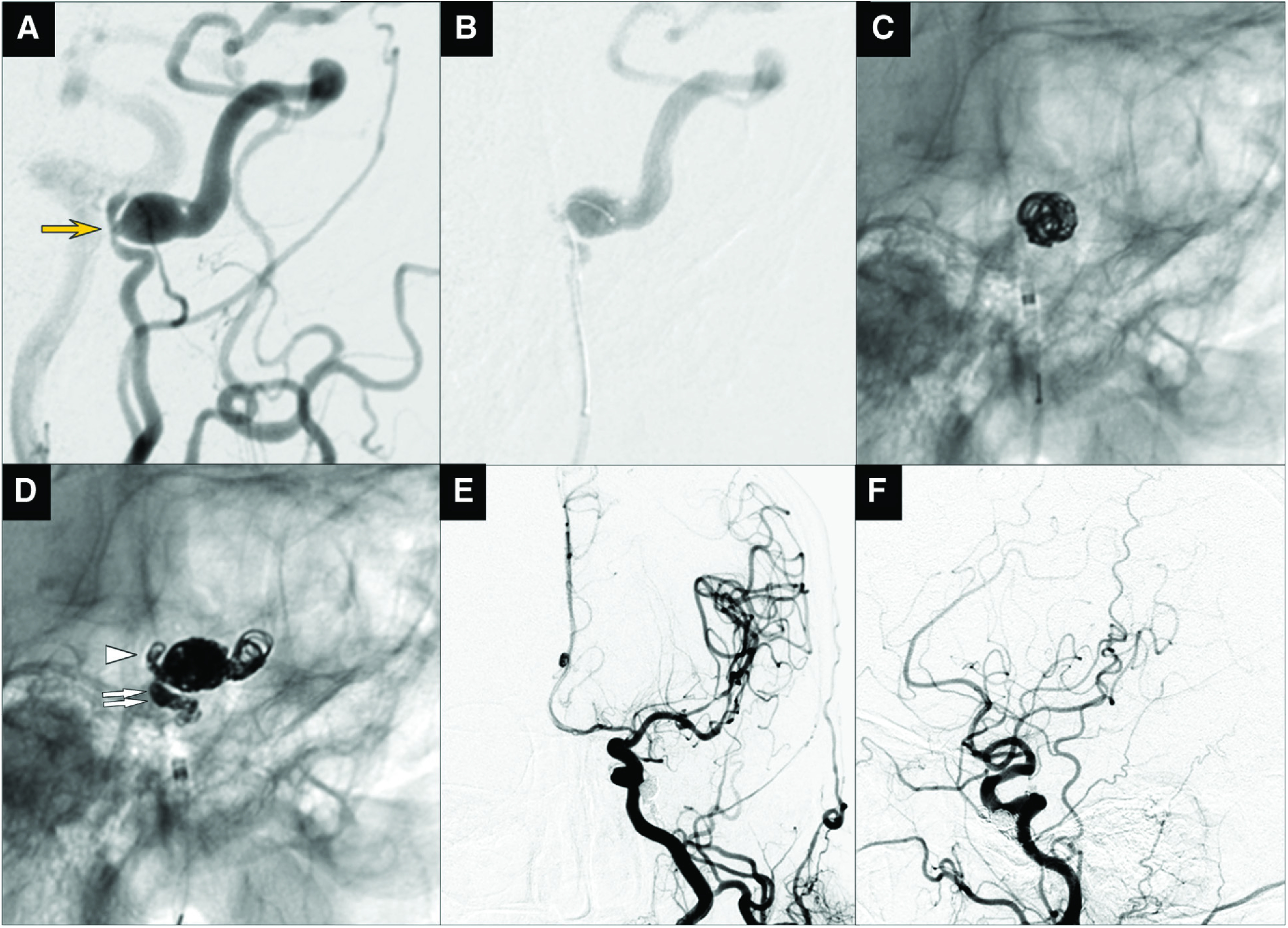
Case presentation: A 78-year-old woman was admitted to our hospital with loss of consciousness, right hemiparesis, and motor aphasia. CT demonstrated intracerebral hematoma in the left frontal lobe and subarachnoid hemorrhage. On CTA and MRA, a DAVF was found in the left laterocavernous sinus region associated with the accessory meningeal artery (AMA) and draining directly into the superficial middle cerebral vein. The diagnosis was confirmed by DSA, which revealed a DAVF fed by the large and straight AMA and the internal carotid artery's meningohypophyseal trunk. Endovascular transarterial venous coil embolization was performed through the AMA. A microcatheter was advanced beyond the shunt point into the origin of the draining vein, and coils were placed in the venous and arterial sides of the fistula. The fistula was completely occluded, and 15-month follow-up angiography demonstrated stable obliteration of the fistula.

Conclusion: Transarterial venous coil embolization may be a treatment option for non-sinus-type laterocavernous sinus DAVF with a large fistula size and a large and straight feeding artery.

Keywords: coil; endovascular therapy; middle fossa dural arteriovenous fistula; sphenoid wing; transarterial venous embolization.


Conflict of interest statement

The authors declare no conflicts of interest.

Figures

Fig. 1
Fig. 1. (A) Cerebral hemorrhage was noted in the left frontal lobe on CT at the first examination. (B) DAVF (dashed circle) with a shunt point (arrow) inferior posterior to the left cavernous sinus was noted on 3D-CTA. Blood directly flowed into the superficial middle cerebral vein, and it was accompanied by a varix (arrowhead: drainer). (CE): Similar findings were noted on MRA (arrow: shunt point, arrowhead: drainer). DAVF: dural arteriovenous fistula
Fig. 2
Fig. 2. (AL) On the preoperative 3D-CTA axial source image, the left AMA (black arrowhead: proximal side, white arrowhead: distal side) formed a direct shunt with the superficial middle cerebral vein (double white arrow) in the yellow arrow region after passing through the oval foramen (FO). AMA: accessory meningeal artery; C4: internal carotid artery C4 segment; C5: internal carotid artery C5 segment; FO: foramen ovale
Fig. 3
Fig. 3. Preoperative DSA. (A and B) The fistula was diagnosed as left laterocavernous sinus DAVF in the frontal view (A) and lateral view (B) on left external carotid arteriography. The feeder was the AMA. It directly flowed into the superficial middle cerebral vein, forming cortical venous reflux, and was then accompanied by a varix and flowed out to the superior sagittal sinus thorough the frontal lobe surface. (C) On right anterior oblique 3D-RA, the AMA diameter immediately before the shunt point (arrow) was 1 mm and the diameter was 1.5 mm (arrowhead: varix). (D and E): A feeder from the left internal carotid artery meningohypophyseal trunk was present in the frontal view (D) and lateral view (E) on angiography of the affected side (left) of the internal carotid artery. AMA: accessory meningeal artery; DAVF: dural arteriovenous fistula; RA: rotational angiography
Fig. 4
Fig. 4. Intraoperative findings. (A) Preoperative left external carotid arteriography, right anterior oblique view (arrow: shunt point). The distal AMA served as a joint with the feeder from the internal carotid artery’s meningohypophyseal trunk. (B) The microcatheter was guided to the drainer side beyond the shunt point through the AMA. (C and D) Coil occlusion was applied in the order from the origin of the drainer (C) to the distal AMA (D, arrowhead) and proximal AMA (D, double arrow). (E and F) On postoperative left common carotid arteriography, the DAVF disappeared in the frontal view (E) and lateral view (F). AMA: accessory meningeal artery

Similar articles

Cited by

References

    1. Nakamura H, Sase T, Wakui D, et al. . Non sinus type dural arteriovenous fistula of the middle cranial fossa: a report of two cases. Surg Cereb Stroke 2016; 44: 151–156. (in Japanese)
    1. Ghali MGZ. Sphenoid dural arteriovenous fistulas. Neurosurg Rev 2021; 44: 77–96. - PubMed
    1. Hiramatsu M, Sugiu K, Haruma J, et al. . Dural arteriovenous fistulas in the parasellar region other than the cavernous sinus. JNET J Neuroendovasc Ther 2020; 14: 593–604. - PMC - PubMed
    1. Baik SK, Kim YW, Lee SW, et al. . A treatment option for nontraumatic adult-type dural arteriovenous fistulas: transarterial venous coil embolization. World Neurosurg 2014; 82: 417–422. - PubMed
    1. Fukai J, Terada T, Kuwata T, et al. . Transarterial intravenous coil embolization of dural arteriovenous fistula involving the superior sagittal sinus. Surg Neurol 2001; 55: 353–358. - PubMed

Publication types