. 2023 Jul 12:17:1115385.
doi: 10.3389/fncel.2023.1115385. eCollection 2023.

Effect of isolated intracranial hypertension on cerebral perfusion within the phase of primary disturbances after subarachnoid hemorrhage in rats

Affiliations

Effect of isolated intracranial hypertension on cerebral perfusion within the phase of primary disturbances after subarachnoid hemorrhage in rats

Guangshan Hao et al. Front Cell Neurosci. .

Abstract

Introduction: Elevated intracranial pressure (ICP) and blood components are the main trigger factors starting the complex pathophysiological cascade following subarachnoid hemorrhage (SAH). It is not clear whether they independently contribute to tissue damage or whether their impact cannot be differentiated from each other. We here aimed to establish a rat intracranial hypertension model that allows distinguishing the effects of these two factors and investigating the relationship between elevated ICP and hypoperfusion very early after SAH.

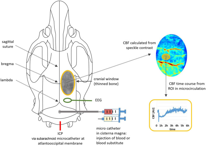
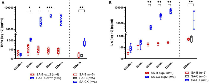
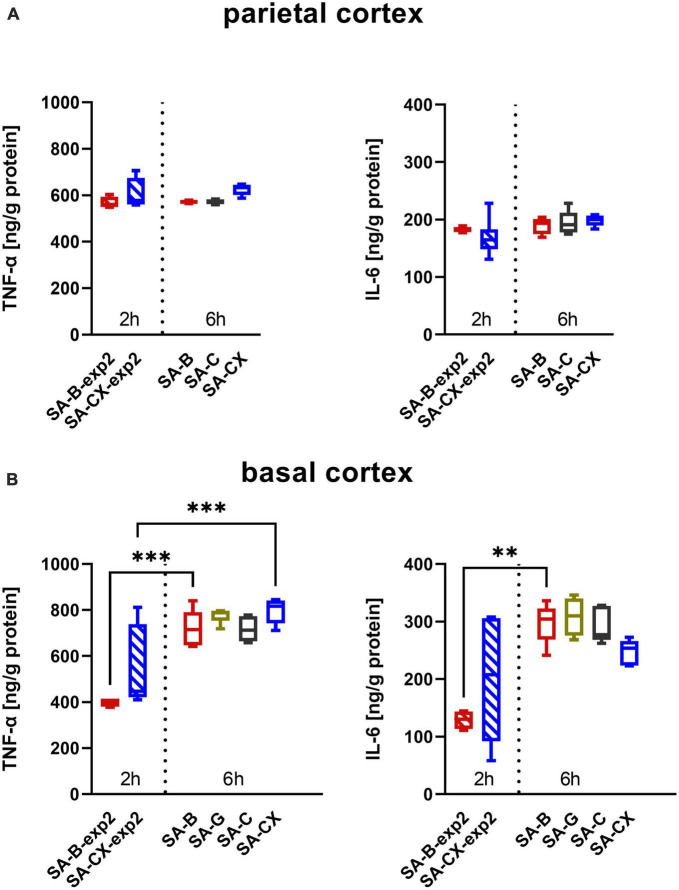
Methods: Blood or four different types of fluids [gelofusine, silicone oil, artificial cerebrospinal fluid (aCSF), aCSF plus xanthan (CX)] were injected into the cisterna magna in anesthetized rats, respectively. Arterial blood pressure, ICP and cerebral blood flow (CBF) were continuously measured up to 6 h after injection. Enzyme-linked immunosorbent assays were performed to measure the pro-inflammatory cytokines interleukin 6 (IL-6) and tumor necrosis factor α (TNF-α) in brain cortex and peripheral blood.

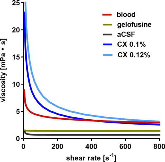
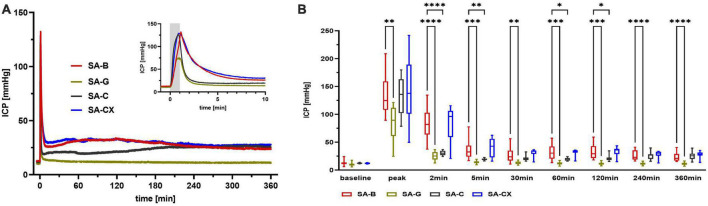
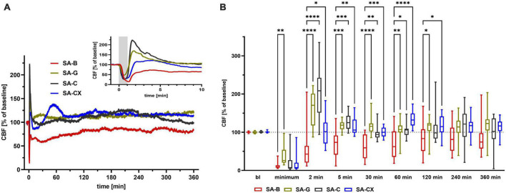
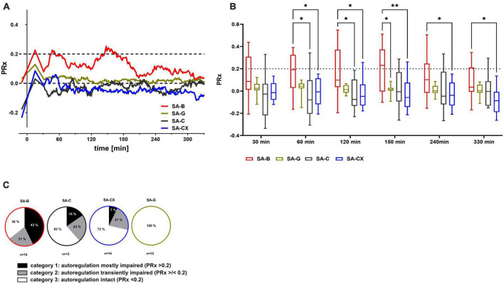
Results: Silicone oil injection caused deaths of almost all animals. Compared to blood, gelofusine resulted in lower peak ICP and lower plateau phase. Artificial CSF reached a comparable ICP peak value but failed to reach the ICP plateau of blood injection. Injection of CX with comparable viscosity as blood reproduced the ICP course of the blood injection group. Compared with the CBF course after blood injection, CX induced a comparable early global ischemia within the first minutes which was followed by a prompt return to baseline level with no further hypoperfusion despite an equal ICP course. The inflammatory response within the tissue did not differ between blood or blood-substitute injection. The systemic inflammation was significantly more pronounced in the CX injection group compared with the other fluids including blood.

Discussion: By cisterna magna injection of blood substitution fluids, we established a subarachnoid space occupying rat model that exactly mimicked the course of ICP in the first 6 h following blood injection. Fluids lacking blood components did not induce the typical prolonged hypoperfusion occurring after blood-injection in this very early phase. Our study strongly suggests that blood components rather than elevated ICP play an important role for early hypoperfusion events in SAH.

Keywords: animal model; cerebral autoregulation; cerebral blood flow; early brain injury; inflammation; intracranial hypertension; rat; subarachnoid hemorrhage.


Conflict of interest statement

The authors declare that the research was conducted in the absence of any commercial or financial relationships that could be construed as a potential conflict of interest.

Figures

FIGURE 1
FIGURE 1
Illustration of the experimental setup: positions of microcatheters for fluid injection, for ICP monitoring and for EEG electrode, cranial window with thinned bone, image processing for time course analysis.
FIGURE 2
FIGURE 2
Overview on experimental group allocation and drop out numbers: (A) experimental study 1; (B) experimental study 2.
FIGURE 3
FIGURE 3
Dynamic viscosity of fluids: whereas aCSF and gelofusine show a low dynamic viscosity, aCSF with 0.1% xanthan (CX) best resembles that of blood.
FIGURE 4
FIGURE 4
ICP: (A) ICP courses induced by the fluids throughout the measurement of 6 h, insert: first 10 min enlarged (gray area depicting the 1 min fluid injection period). (B) Quantitative analysis at distinct time points (****p < 0.0001; ***p < 0.001; **p < 0.01; *p < 0.05).
FIGURE 5
FIGURE 5
CBF: (A) CBF courses induced by the fluids throughout the measurement of 6 h, insert: first 10 min enlarged (gray area depicting the 1 min fluid injection period). (B) Quantitative analysis at distinct time points (****p < 0.0001; ***p < 0.001; **p < 0.01; *p < 0.05).
FIGURE 6
FIGURE 6
PRx as measure for cerebral autoregulation following fluid injection: the pressure reactivity index PRx, for assessment of cerebral autoregulation (CA) was calculated as the continuous Pearson correlation coefficient between intracranial pressure and arterial blood pressure at low frequencies. PRx > 0.2 indicates impaired CA, whereas PRx < 0.2 points toward an intact CA. (A) Median of PRx time courses for all groups. (B) Quantitative analysis at distinct time points (**p < 0.01; *p < 0.05). (C) Distribution between categories of mostly (more than 50% of the datapoints are > 0.2) or transiently (less than 50% but more than 0% of all datapoints are > 0.2) impaired or intact (0% of the datapoints are > 0.2, means all datapoints are < 0.2) CA according to the cutoff value of PRx 0.2 (% of animals in each group).
FIGURE 7
FIGURE 7
Systemic inflammatory response assessed in blood samples: (A) TNF-α, (B) IL-6. Data from experimental study 2, with samples taken every 30 min up to 2 h after blood or CX-injection (SA-B-exp2; SA-CX-exp2) are arranged together with data from experimental study 1, with samples taken at the end of the 6 h observation period (SA-B; SA-C; SA-CX) (***p < 0.001; **p < 0.01; *p < 0.05).
FIGURE 8
FIGURE 8
Inflammatory response from brain samples: (A) parietal and (B) basal cortex assessed by TNF-α (left) and IL-6 (right) analysis: data from experimental study 2, with samples taken at 2 h after blood or CX-injection (SA-B-exp2; SA-CX-exp2) are arranged together with data from experimental study 1, with samples taken at the end of the 6 h observation period (SA-B; SA-C; SA-CX) (***p < 0.001; **p < 0.01).

Similar articles

Cited by

References

    1. Abraham M. K., Chang W. W. (2016). Subarachnoid hemorrhage. Emerg. Med. Clin. North Am. 34 901–916. 10.1016/j.emc.2016.06.011 - DOI - PubMed
    1. Amico C., Tornetta T., Scifo C., Blanco A. R. (2015). Antioxidant effect of 0.2% xanthan gum in ocular surface corneal epithelial cells. Curr. Eye Res. 40 72–76. 10.3109/02713683.2014.914542 - DOI - PubMed
    1. Andereggen L., Neuschmelting V., von Gunten M., Widmer H. R., Takala J., Jakob S. M., et al. (2014). The rabbit blood-shunt model for the study of acute and late sequelae of subarachnoid hemorrhage: Technical aspects. J. Vis. Exp. 92:e52132. 10.3791/52132 - DOI - PMC - PubMed
    1. Ascenzi P., Bocedi A., Visca P., Altruda F., Tolosano E., Beringhelli T., et al. (2005). Hemoglobin and heme scavenging. IUBMB Life 57 749–759. 10.1080/15216540500380871 - DOI - PubMed
    1. Bach A., Conzen C., Schubert G. A., Bleilevens C., Lindauer U. (2019). Acute changes of pro-inflammatory markers and corticosterone in experimental subarachnoid haemorrhage: A prerequisite for severity assessment. PLoS One 14:e0220467. 10.1371/journal.pone.0220467 - DOI - PMC - PubMed

LinkOut - more resources