

Facial nerve schwannoma: Case report and brief review of the literature
- PMID: 37502483
- PMCID: PMC10369394
- DOI: 10.1016/j.radcr.2023.06.043
Facial nerve schwannoma: Case report and brief review of the literature
Abstract
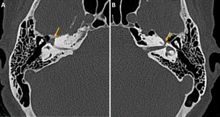
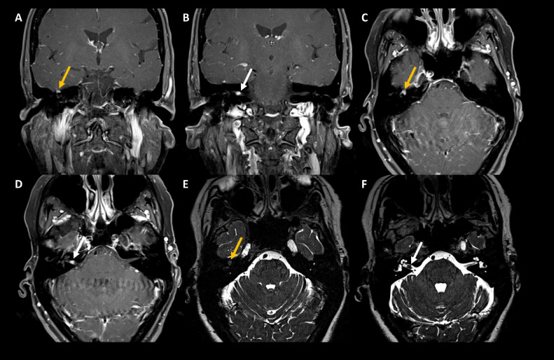
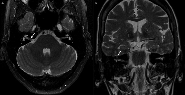
Schwannomas are rare nerve sheath tumors that can occur throughout the body, and are symptomatic based on location, size, and impingement on adjacent structures. These tumors are often benign lesions and occur sporadically or from genetic conditions such as neurofibromatosis. Schwannomas may arise from peripheral nerves, gastrointestinal nerves, spinal nerve roots and cranial nerves. Facial nerve schwannomas arise from cranial nerve VII, commonly involving the geniculate ganglion, labyrinthine segment, and internal auditory canal. While small lesions are asymptomatic, larger lesions can cause facial nerve paralysis, and facial spasms. Lesions in the internal auditory canal can cause hearing loss, tinnitus, vertigo, and otalgia. High-resolution CT imaging and MRI imaging are useful for distinguishing between other pathologies that arise from the same region. High-resolution CT scans can show bony degeneration of nearby structures such as the labyrinth or ossicles. MRI imaging shows hypo intensity on T1 imaging, and hyperintensity on T2 imaging. On T1 postcontrast, enhancement can be homogenous or heterogeneous with cystic degeneration if the lesion is large. Nodular enhancement is commonly seen on facial nerve schwannomas within the internal auditory canal. Vestibular schwannomas involving CN VIII are more common, and appear similar to facial nerve schwannomas, but can be distinguished apart due to growth in the geniculate ganglion and/or the labyrinthine segment. Management of asymptomatic or mild lesions is typically conservative with follow up imaging, and surgery for larger lesions. Here, we present a case of a facial nerve schwannoma in a 57-year-old woman.
Keywords: Facial nerve palsy; Facial nerve schwannoma; Geniculate ganglion; Internal auditory canal; MRI.
© 2023 Published by Elsevier Inc. on behalf of University of Washington.
Figures
References
-
- Beaman F.D., Kransdorf M.J., Menke D.M. Schwannoma: radiologic-pathologic correlation. Radiographics. 2004;24(5):1477–1481. - PubMed
-
- Hassell D.S., Bancroft L.W., Kransdorf M.J., Peterson J.J., Berquist T.H., Murphey M.D., et al. Imaging appearance of diffuse neurofibroma. American Journal of Roentgenology. 2008;190(3):582–588. - PubMed
-
- National Cancer Institute. Schwannoma. 2020 Available at: https://www.cancer.gov/pediatric-adult-rare-tumor/rare-tumors/rare-soft-....
-
- Schmidt R.F., Boghani Z., Choudhry O.J., Eloy J.A., Jyung R.W. Incidental vestibular schwannomas: a review of prevalence, growth rate, and management challenges. Neurosurg Focus. 2012;33(3):E4. - PubMed
-
- Skolnik A.D., Loevner L.A., Sampathu D.M., Newman J.G., Lee J.Y., Bagley L.J., et al. Cranial nerve schwannomas: diagnostic imaging approach. Radiographics. 2016;36(5):1463–1477. - PubMed
Publication types
LinkOut - more resources
Full Text Sources
Research Materials
