

Case Reports
doi: 10.1016/j.radcr.2023.07.029.
eCollection 2023 Oct.
A case of intrascrotal extratesticular schwannoma
Affiliations
- PMID: 37502485
- PMCID: PMC10369397
- DOI: 10.1016/j.radcr.2023.07.029
Item in Clipboard
Case Reports
A case of intrascrotal extratesticular schwannoma
Radiol Case Rep.
.
Abstract
Schwannomas are benign tumors arising from Schwann cells, which compose the myelin sheath covering peripheral nerves. Although schwannomas can develop in various locations throughout the human body, the scrotum is a rare site for development of a schwannoma. Furthermore, to the best of our knowledge, no study to date has focused on the detailed imaging findings of intrascrotal schwannoma.
Keywords: Magnetic resonance imaging; Schwannoma; Scrotal tumor; Tunica albuginea.
© 2023 The Authors. Published by Elsevier Inc. on behalf of University of Washington.
Figures
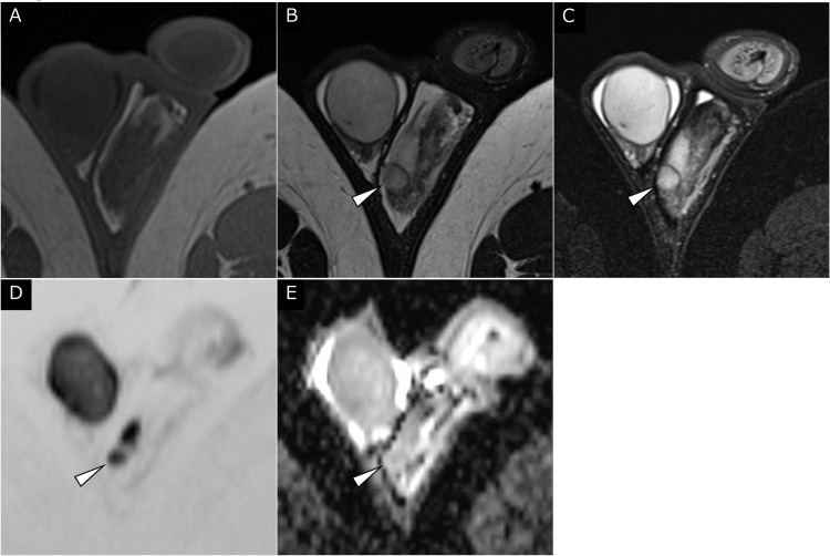
Axial MRI of the left scrotal mass. (A) T1WI failed to demonstrate the tumor. A well-defined slightly inhomogeneous 10-mm mass of high signal intensity (arrowheads) was present at the periphery of the left testis on (B) T2WI and (C) fat-suppressed T2WI. (D, E) On DWI, the mass (arrowheads) showed diffusion restriction comparable to that of the normal testis (apparent diffusion coefficient of the mass: 1.1 × 10⁻³ mm2/s).

Coronal T2WI of the left scrotal mass. (A–C) A small mass with a rim of low signal intensity (arrowheads) was depicted between the left testis (T) and epididymis (E). The low-signal-intensity rim had continuity with the tunica albuginea and vaginalis (arrows). The mass showed similar or slightly lower signal intensity compared with the normal testis.
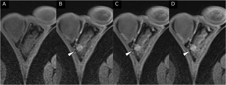
Dynamic contrast-enhanced MRI, axial images. (A) Precontrast fat-suppressed T1WI failed to demonstrate the tumor. The mass showed inhomogeneous contrast enhancement on postcontrast scans at (B) 40 seconds, (C) 70 seconds, and (D) 150 seconds after intravenous gadolinium injection.

Histological section (hematoxylin and eosin stain) of the scrotal tumor. (A) Pathologically, the tumor (*) showed continuity with the tunica albuginea (TA) and was adjacent to the left testis (T). (B) No abnormal findings were observed in the left testis and epididymis.
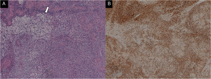
Histological sections (hematoxylin and eosin stain and immunostaining for S-100) of the scrotal tumor. (A) On hematoxylin and eosin staining, the tumor consisted of spindle cell proliferation without an increase in mitotic figures. No intratumoral hemorrhage or necrosis was observed. A component consisting of spindle cells with a loose myxoid matrix (arrow) was partially confirmed. (B) The tumor cells showed intense immunoreactivity for S-100 staining. A paratesticular schwannoma was pathologically confirmed.
References
-
- MacCollin M, Chiocca EA, Evans DG, Friedman JM, Horvitz R, Jaramillo D, et al. Diagnostic criteria for schwannomatosis. Neurology. 2005;64(11):1838–1845. doi: 10.1212/01.WNL.0000163982.78900.AD. - DOI - PubMed
Publication types
LinkOut - more resources
Full Text Sources
