Case Reports
. 2023 Jul 22;18(10):3434-3437.
doi: 10.1016/j.radcr.2023.07.005. eCollection 2023 Oct.

A rare cause of right-upper quadrant abdominal pain: Epiploic appendagitis of the hepatic flexure

Affiliations
Case Reports

A rare cause of right-upper quadrant abdominal pain: Epiploic appendagitis of the hepatic flexure

Vincenzo Cirimele et al. Radiol Case Rep. .

Abstract

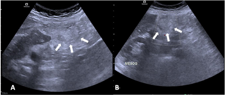
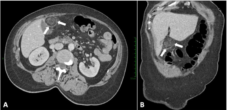
Epiploic appendagitis (EA) is an uncommon cause of acute abdominal pain that may mimic other causes of acute abdomen. Epiploic appendages are outpouching of fat tissue located on the external wall of the colon, being more numerous in the descending and sigmoid colon that account for up to 80% of EA cases. We present the case of a 59-year-old woman with right upper quadrant pain. Abdominal ultrasound and contrast-enhanced computed tomography suggested the diagnosis of epiploic appendagitis of the right colonic flexure. Our case highlights the fact that epiploic appendagitis may occur in unusual locations and must be included in the differential diagnosis of acute abdominal pain, in order to avoid unnecessary medical and surgical treatment.

Keywords: Acute abdomen; Computed tomography; Emergency radiology; Epiploic appendagitis; Ultrasound.


Figures

Fig 1
Fig. 1
Abdominal US showing a hyperechoic rounded mass in the right hypocondrium adjacent to the lower margin of IVb hepatic segment close to the ascending colon (white arrows in A and B). The mass is surrounded by a hypoechoic rim. Color Doppler US shows absent internal blood flow (B).
Fig 2
Fig. 2
Axial (A) and coronal (B) contrast-enhanced CT demonstrating a formation with heterogeneous adipose matrix and peripheral dense rim (white arrows in A and B) adjacent to the right colic flexure. The findings were consistent with epiploic appendagitis.

Similar articles

References

    1. Boardman J, Kaplan KJ, Hollcraft C, Bui-Mansfield LT. Radiologic-pathologic conference of Keller Army Community Hospital at West Point, the United States Military Academy: torsion of the epiploic appendage. AJR Am J Roentgenol. 2003;180(3):748. doi: 10.2214/ajr.180.3.1800748. - DOI - PubMed
    1. Gaur S, Jawahar RP, Prasad R, Prabakaran M. Epiploic appendagitis - a rare cause of acute lower abdominal pain. Radiol Case Rep. 2021;16(5):1144–1147. doi: 10.1016/j.radcr.2021.02.026. Erratum in: Radiol Case Rep. 2022 Sep 29;17(12):4933. PMID: 33747330; PMCID: PMC7960494. - DOI - PMC - PubMed
    1. Singh AK, Gervais DA, Hahn PF, Sagar P, Mueller PR, Novelline RA. Acute epiploic appendagitis and its mimics. Radiographics. 2005;25(6):1521–1534. doi: 10.1148/rg.256055030. - DOI - PubMed
    1. Almeida AT, Melão L, Viamonte B, Cunha R, Pereira JM. Epiploic appendagitis: an entity frequently unknown to clinicians–diagnostic imaging, pitfalls, and look-alikes. AJR Am J Roentgenol. 2009;193(5):1243–1251. doi: 10.2214/AJR.08.2071. - DOI - PubMed
    1. Trovato P, Simonetti I, Verde F, Lomoro P, Vinci G, Tarotto L, et al. Acute epiploic appendagitis: ultrasound and computed tomography findings of a rare case of acute abdominal pain and the role of other imaging techniques. Pol J Radiol. 2020;85:e178–e182. doi: 10.5114/pjr.2020.94335. - DOI - PMC - PubMed

Publication types

LinkOut - more resources