. 2023 Jul 18:29:230-240.
doi: 10.1016/j.bioactmat.2023.07.002. eCollection 2023 Nov.

C-176 loaded Ce DNase nanoparticles synergistically inhibit the cGAS-STING pathway for ischemic stroke treatment

Affiliations

C-176 loaded Ce DNase nanoparticles synergistically inhibit the cGAS-STING pathway for ischemic stroke treatment

Zhixin Zhu et al. Bioact Mater. .

Abstract

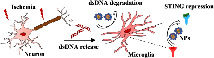
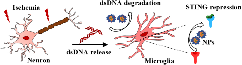
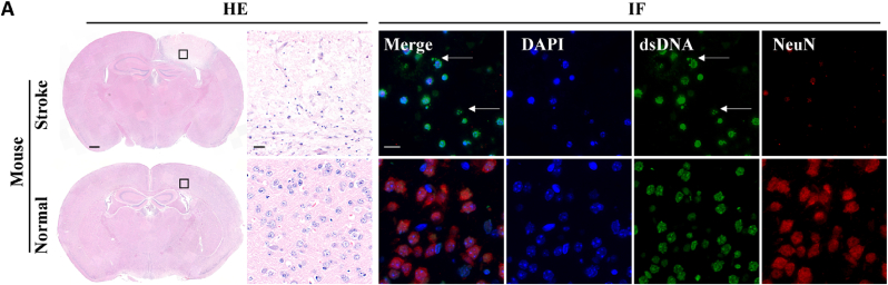
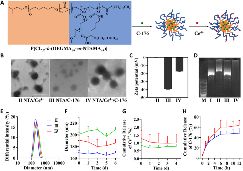
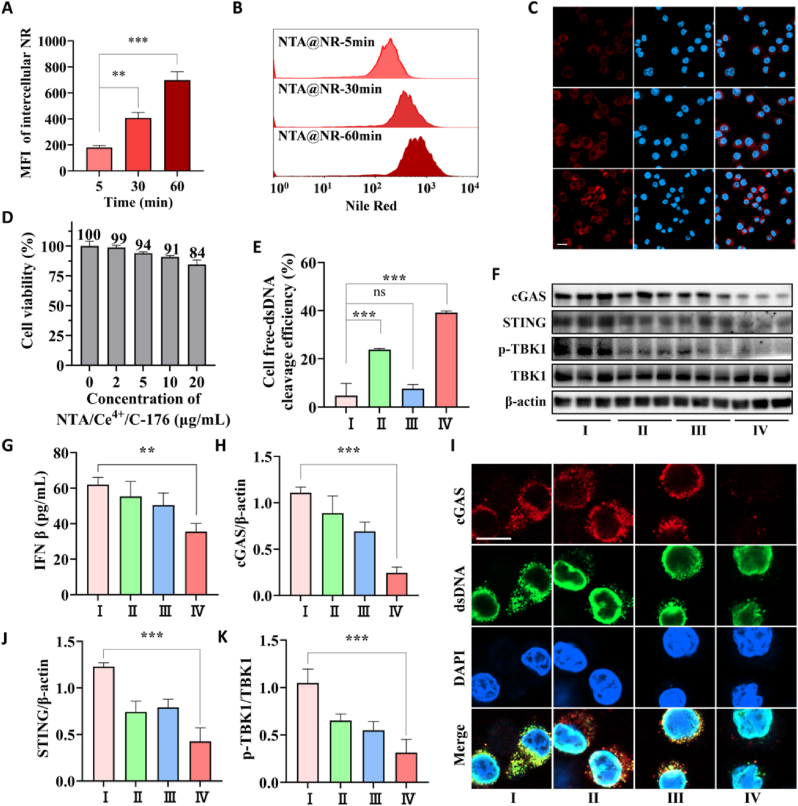
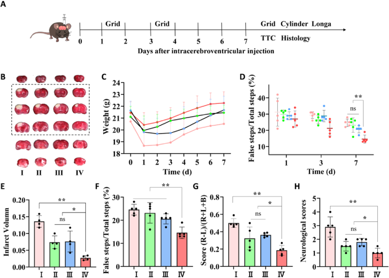
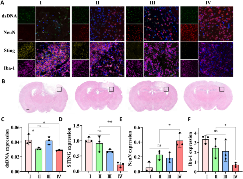
The neuroinflammatory responses following ischemic stroke cause irreversible nerve cell death. Cell free-double strand DNA (dsDNA) segments from ischemic tissue debris are engulfed by microglia and sensed by their cyclic GMP-AMP synthase (cGAS), which triggers robust activation of the innate immune stimulator of interferon genes (STING) pathway and initiate the chronic inflammatory cascade. The decomposition of immunogenic dsDNA and inhibition of the innate immune STING are synergistic immunologic targets for ameliorating neuroinflammation. To combine the anti-inflammatory strategies of STING inhibition and dsDNA elimination, we constructed a DNase-mimetic artificial enzyme loaded with C-176. Nanoparticles are self-assembled by amphiphilic copolymers (P[CL35-b-(OEGMA20.7-co-NTAMA14.3)]), C-176, and Ce4+ which is coordinated with nitrilotriacetic acid (NTA) group to form corresponding catalytic structures. Our work developed a new nano-drug that balances the cGAS-STING axis to enhance the therapeutic impact of stroke by combining the DNase-memetic Ce4+ enzyme and STING inhibitor synergistically. In conclusion, it is a novel approach to modulating central nervus system (CNS) inflammatory signaling pathways and improving stroke prognosis.

Keywords: Anti-inflammation; Ce-based nano-nuclease; Ischemic stroke; The cGAS-STING signaling pathway.


Conflict of interest statement

The authors affirm that they have no known financial or interpersonal conflicts that could have appeared to have an impact on the research presented in this study.

Figures

Image 1
Graphical abstract
Scheme 1
Scheme 1
The therapeutic mechanism of NTA/Ce4+/C-176 nanoparticles.
Fig. 1
Fig. 1
The representative images of the HE staining, NeuN (red)/dsDNA (green) coimmunostaining of the MCAO model mice and corresponding controls. Scale bars: 500 μm and 20 μm for HE staining and 20 μm for immunostaining. The dsDNA displayed cell-free dsDNA (green) as highlighted with white arrows.
Fig. 2
Fig. 2
The characterization of NTA/Ce4+/C-176 nanoparticles. (A) Schematic illustration of amphophilic block copolymer P[CL35-b-(OEGMA20.7-co-NTAMA14.3)] structure and preparation of C-176-loaded nanoparticles coordinated with Ce4+. (B) TEM images of NTA/Ce4+, NTA/C-176, and NTA/Ce4+/C-176 freeze-dried in water; scale bar = 200 nm. (C) Zeta potential of NTA/Ce4+, NTA/C-176, NTA/Ce4+/C-176 in water (n = 3). (D) Agarose gel (1%) electrophoresis showing plasmids PUC18 (2686 bp) cleavage by Ce(NH4)2(NO3)6, NTA/Ce4+, NTA/C-176, NTA/Ce4+/C-176. Lane M: DNA marker; lane Ⅰ: PUC18 with Ce(NH4)2(NO3)6; lane Ⅱ: PUC18 with NTA/Ce4+; lane Ⅲ: PUC18 with NTA/C-176; lane Ⅳ: PUC18 with NTA/Ce4+/C-176. (E) The differential intensity of NTA/Ce4+, NTA/C-176, NTA/Ce4+/C-176. (F) Average hydrodynamic diameters of NTA/Ce4+, NTA/C-176, NTA/Ce4+/C-176 in water at 0, 1, 2, 5, 6 d (n = 3). (G) Cumulative release of Ce4+ from NTA/Ce4+ and NTA/Ce4+/C-176 at 0, 1, 2, 3, 4 d. The Ce signals were monitored by ICP-MS (n = 3). (H) Cumulative release of C-176 from NTA/C-176 and NTA/Ce4+/C-176 at 0, 1, 2, 4, 6, 8, 10, 12 h. The C-176 signals were monitored by HPLC (n = 6).
Fig. 3
Fig. 3
Biocompatibility and catalytic activity of NTA/Ce4+/C-176 in vitro. (A) Quantified NTA/Ce4+/Nile red cellular uptake in BV2 cells by flow cytometry analysis with increasing co-incubation time (n = 3). Data are presented as mean ± SD. **P < 0.01, ***P < 0.001. (B) Typical flow cytometry data of cellular uptake of NTA/Ce4+/Nile red in BV2 cells. (C) Confocal images of intracellular NTA/Ce4+/Nile red in BV2 cells. NTA/Ce4+/Nile red indicated by a red signal and nuclei stained by DAPI indicated by a blue signal; scale bar = 20 μm. (D) Viability of BV2 cells treated for 24 h with various concentrations of NTA/Ce4+/C-176 (n = 3). (E) Cell-free dsDNA cleavage efficiency of the pico-green-stained BV2-cultured supernatant. Data are means ± SEM (n = 3 independent experiments; ***P < 0.001 by one-way ANOVA with Tukey's multiple comparison tests, ns means no significant difference). (F) Representative immunoblotting of cGAS, STING, p-TBK1, and TBK1 in BV2 after indicated treatments (n = 3), using β-Actin as control. ELISA of (G) IFNβ, blotting quantification of (H) cGAS, (J) STING, and (K) p-TBK1. (I) Cellular immunofluorescence images of dsDNA (green) and cGAS (red) in BV2 cells. Scar bar = 10 μm.
Fig. 4
Fig. 4
Effect of NTA/Ce4+/C-176 on brain infarct volume and functional motor recovery after stroke. (A) The experimental timeline. (B) Representative images of the TTC-stained brain slice after 7 d post-stroke. The normal tissue was stained red, while the injured tissue was unstained white. (C) Body weight before cerebral infarction (d 0) and after cerebral infarction with different treatments (n = 5). (D) Grid test on 1, 3, and 7 d after stroke. (n = 5). (E) Infarct volume of the TTC-staining at 7 d post-stroke (n = 4). (F) Grid test on 7 d after stroke (n = 5). (G) Cylinder test on 7 d after therapy (n = 5). (H) Neurological scores on 3 d after therapy (n = 5). *P < 0.05, **P < 0.01, ns means no significant difference.
Fig. 5
Fig. 5
NTA/Ce4+/C-176 decreased neuroinflammation and increased neurogenesis in vivo. (A) Immunofluorescence of dsDNA (injury markers, green), NeuN (neuron markers, red), DAPI (nucleus markers, blue), STING (injury markers, yellow), Iba-1 (microglia markers, purple). The white arrows indicated dsDNA-positive area (injury); scale bar = 20, 40 μm). (B) The corresponding representative immunohistochemical brain slices of the immunofluorescence pictures (scale bar = 500 μm). (C) Quantitative analysis of relative dsDNA levels (n = 3), STING levels in (D) (n = 4). NeuN levels in (E) and Iba-1 in (F) (n = 3) normalized to nuclei, respectively. *P < 0.05, **P < 0.01, ns means no significant difference.
Fig. 6
Fig. 6
Toxicological evaluations after 7 d of treatment via different injections. (A–D) Blood biochemical values of blood urea nitrogen (BUN), aspartate aminotransferase (AST), alanine aminotransferase (ALT), and creatinine (CRE) at 7 d post-injection (n = 4). (E–H) Routine blood indexes of red blood cell (RBC), platelets (PLT), neutrophil (Neu), and lymphocytes (Lym) at 7 d post-injection. ns means no significant difference. (I) H&E staining of heart, liver, spleen, lung, kidney, and brain at 7 d post-injection; scar bar = 20 μm.

References

    1. Wu S., Wu B., Liu M., et al. Stroke in China: advances and challenges in epidemiology, prevention, and management. Lancet Neurol. 2019;18(4):394–405. - PubMed
    1. Tian X., Fan T., Zhao W., et al. Recent advances in the development of nanomedicines for the treatment of ischemic stroke. Bioact. Mater. 2021;6(9):2854–2869. - PMC - PubMed
    1. Fu Y., Liu Q., Anrather J., et al. Immune interventions in stroke. Nat. Rev. Neurol. 2015;11(9):524–535. - PMC - PubMed
    1. Shi K., Tian D.-C., Li Z.-G., et al. Global brain inflammation in stroke. Lancet Neurol. 2019;18(11):1058–1066. - PubMed
    1. Salter M.W., Stevens B. Microglia emerge as central players in brain disease. Nat. Med. 2017;23(9):1018–1027. - PubMed