[Preprint]. 2023 Jul 11:rs.3.rs-3146716.
doi: 10.21203/rs.3.rs-3146716/v1.

Selective Blood Cell Hitchhiking in Whole Blood with Ionic Liquid-Coated PLGA Nanoparticles to Redirect Biodistribution After Intravenous Injection

Affiliations

Selective Blood Cell Hitchhiking in Whole Blood with Ionic Liquid-Coated PLGA Nanoparticles to Redirect Biodistribution After Intravenous Injection

Christine M Hamadani et al. Res Sq. .

Abstract

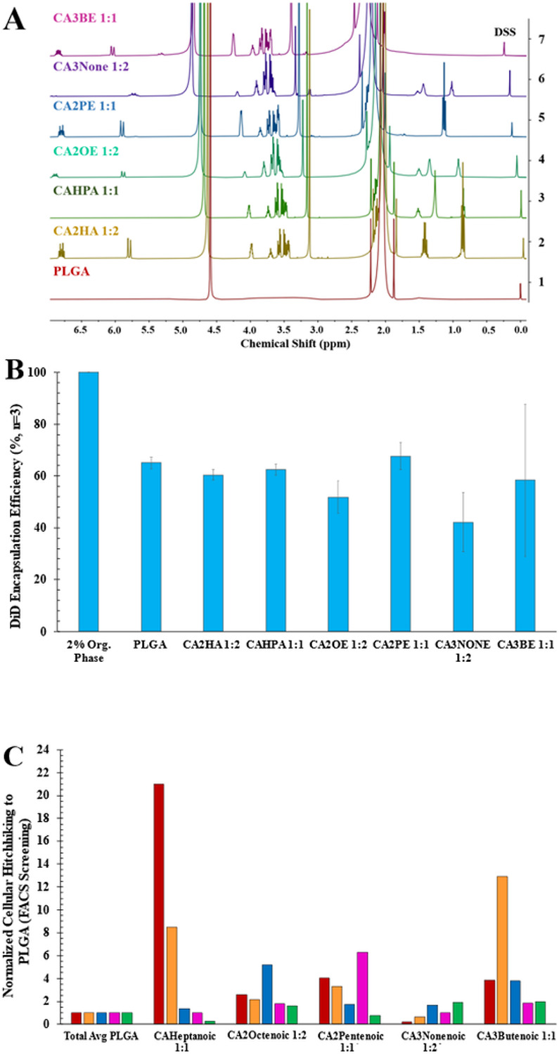
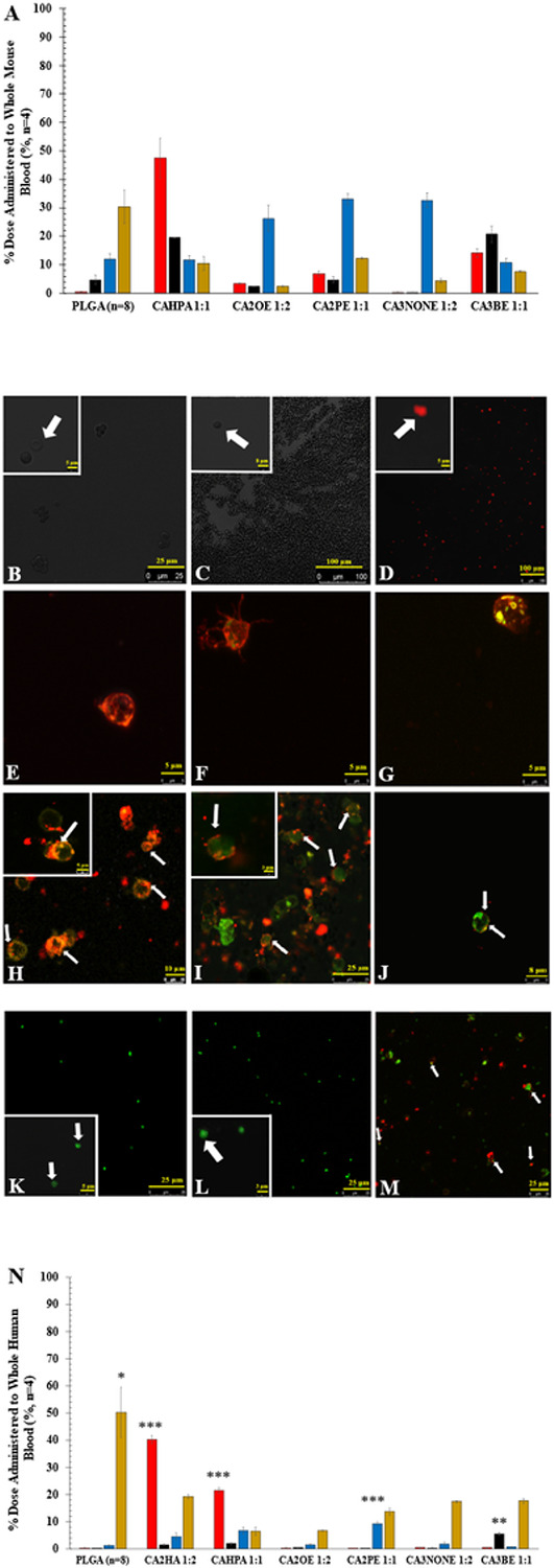
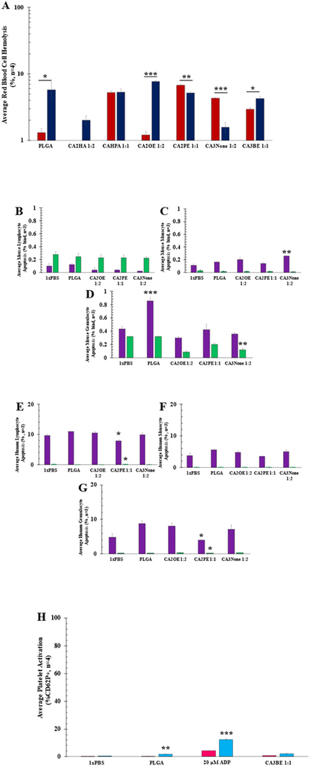
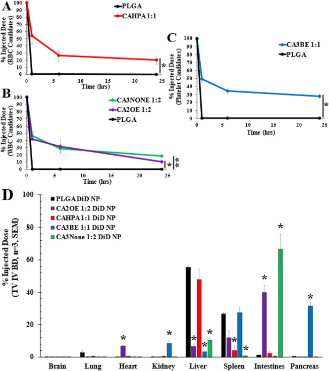
Less than 5% of intravenously-injected nanoparticles (NPs) reach destined sites in the body due to opsonization and immune-based clearance in vascular circulation. By hitchhiking in situ onto specific blood components post-injection, NPs can selectively target tissue sites for unprecedentedly high drug delivery rates. Choline carboxylate ionic liquids (ILs) are biocompatible liquid salts <100X composed of bulky asymmetric cations and anions. This class of ILs has been previously shown to significantly extend circulation time and redirect biodistribution in BALB/c mice post-IV injection via hitchhiking on red blood cell (RBC) membranes. Herein, we synthesized & screened 60 choline carboxylic acid-based ILs to coat PLGA NPs and present the impact of structurally engineering the coordinated anion identity to selectively interface and hitchhike lymphocytes, monocytes, granulocytes, platelets, and RBCs in whole mouse blood for in situ targeted drug delivery. Furthermore, we find this nanoparticle platform to be biocompatible (non-cytotoxic), translate to human whole blood by resisting serum uptake and maintaining modest hitchhiking, and also significantly extend circulation retention over 24 hours in BALB/c healthy adult mice after IV injection. Because of their altered circulation profiles, we additionally observe dramatically different organ accumulation profiles compared to bare PLGA NPs. This study establishes an initial breakthrough platform for a modular and transformative targeting technology to hitchhike onto blood components with high efficacy and safety in the bloodstream post-IV administration.


Conflict of interest statement

COI Statement: EELT and CMH are named as inventors on intellectual property disclosures describing this work

Figures

Figure 1
Figure 1. IL-PLGA DiD NP top candidates demonstrate unique chemical compositions when coating PLGA NPs, varying but functional encapsulation efficiencies (EE, %), and magnitudes-greater hitchhiking affinities towards specific cell-types vs. PLGA NPs in whole blood.
A) Quantitative 1H NMR spectroscopy (DSS, 0.00 ppm: 0.2 mg DSS/1 mg NPs in D2O) illustrates the impact of varying carboxylate anion structure (violet-gold) on the assembly of PLGA NP surfaces (red), quantified in Table S3. B) DiD Encapsulation Efficiency (EE%) by fluorescent plate reader shows the impact of each top candidate IL coating on the fluorescence of PLGA DiD NPs, ranging from 40-70%, when directly compared to the organic phase used for synthesis (Table S4). C) From a FACS screen of 60+ screened IL-PLGA DiD NPs in whole blood (normalized to bare PLGA DiD NPs), top IL candidates show unprecedented and selective cellular hitchhiking to RBCs (red), platelets (orange), lymphocytes (blue), monocytes (pink), and granulocytes (green).
Figure 2
Figure 2. IL-PLGA DiD NP hitchhiking candidates show prominent qualitative and quantitative cellular hitchhiking in whole mouse blood via live-cell confocal microscopy (37 °C; yellow: PE-CDllb+, green: FITC-Ly6G+, red: DiD NPs) and fluorescent plate reader, which translates modestly into human whole blood via generally reduced serum residence (gold) compared to PLGA, and significantly increased fluorescence in RBC (red), platelet (black), and WBC populations (blue).
A) Demonstrates cellular affinity of each top IL-PLGA DiD NP candidate in whole BALB/c mouse blood by fluorescent plate reader. Shown: quantified average % NP distribution per cell type for each top candidate in whole mouse BALB/c blood (n=4, SEM). Color key: Gold (Serum) , Blue (WBC), Black (Platelet), Red (RBC). Pictured by live cell confocal microscopy are the following isolated live cell populations (zoomed-in insets in white boxes) from whole blood treated with: B) 1x PBS-treated RBCs, C) PLGA-treated RBCs, D) CAHPA 1:1-treated RBCs; E) PLGA-treated Lymphocyte WBCs, F) PLGA-treated Dendritic Monocyte WBCs, G) PLGA-treated Granulocyte WBCs; H) CA2OE 1:2-treated Lymphocyte WBCs, I) CA2PE 1:1-treated Monocyte WBCs; J)CA3None 1:2-treated Granulocyte WBCs; K) 1x PBS-treated platelets, L)PLGA-treated platelets, M) CA3BE 1:1-treated platelets. N)Demonstrates translational cellular affinity of each top IL-PLGA DiD NP candidate (n=4) vs. bare PLGA (n=8) in human whole blood. Significance against bare PLGA was performed as a two-tailed t-test of unequal variance, with serum uptake screened across all candidates and bare PLGA via ANOVA (*** = p<0.001, ** = p<0.01, & * = p<0.05).
Figure 3
Figure 3. IL-PLGA NP candidates show high levels of biocompatibility with their respective cellular-hitchhiking affinity-types via RBC hemolysis, WBC apoptosis, and platelet activation assays.
Data represented as average with standard error of mean (SEM). Significance determined by paired two-tailed t-test for two samples at a time between mouse and human samples: *=p<0.05, **=p<0.01, ***=p<0.001. A) Red Blood Cell Hemolysis Assay by fluorescent plate reader of all top candidates and bare PLGA NPs in BALB/c mouse RBCs (red) and Human RBCs (navy) (n=4, SEM). B-G) PI/Annexin V WBC early (purple) vs. late (green) apoptosis assayed by FACS of top lymphocyte (CA2OE 1:2), monocytes (CA2PE 1:1), and granulocyte (CA3None 1:2) hitchhiking candidates from: B-D)whole BALB/c mouse blood or E-G) whole human blood treatment (n=3, ± SEM). H) Platelet activation response by FACS (% activation, CD62P+ from CD41-labeled platelets) to platelet candidate CA3BE 1:1 in whole BALB/c mouse (pink) or human (blue) whole blood (n=4, SEM).
Figure 4
Figure 4. IL-PLGA NP candidates with selective cellular affinity in whole blood significantly extend BALB/c mouse pharmacokinetics (PK) and redirect organ biodistribution (BD) 24 hours after IV-tail-vein injection.
A-C) PK curves & D) BD represented as average (n=3) of injected dose ± SEM. Bare PLGA NPs (black) functionalized at the surface with only carboxylic acid (black) are almost completely cleared from circulation 1 hour after injection and mainly accumulate in the liver & spleen. In contrast, all candidates have significantly higher retention at 24 hours, with WBC candidates bearing similar PK profiles to a PEGylated control. Two-samples evaluated at a time via paired two-tail t-test):*=p<0.05, **=p<0.01. BD: Lymphocyte candidate CA2OE 1:2 (purple) and granulocyte candidate CA3None 1:2 (green) primarily accumulate in the intestine lymphatic vasculature, while platelet candidate CA3BE 1:1 (blue) redirects BD to the pancreas and spleen. RBC candidate CAHPA 1:1 (red) shears selectively in the liver, but with negligible accumulation in the spleen compared to bare PLGA. For BD, * indicates significantly less or greater organ accumulation at p<0.05 relative to bare PLGA NPs (black) per organ type.

Similar articles

References

    1. Palanikumar L., Al-Hosani S., Kalmouni M. et al. pH-responsive high stability polymeric nanoparticles for targeted delivery of anticancer therapeutics. Commun Biol 3, 95 (2020). 10.1038/s42003-020-0817-4 - DOI - PMC - PubMed
    1. Senapati S., Mahanta A.K., Kumar S. et al. Controlled drug delivery vehicles for cancer treatment and their performance. Sig Transduct Target Ther 3, 7 (2018). 10.1038/s41392-017-0004-3 - DOI - PMC - PubMed
    1. Enlow EM, Luft JC, Napier ME, DeSimone JM. Potent engineered PLGA nanoparticles by virtue of exceptionally high chemotherapeutic loadings. Nano Lett. 2011. Feb 9;11(2):808–13. doi: 10.1021/nl104117p. Epub 2011 Jan 25. - DOI - PMC - PubMed
    1. Mitchell M.J., Billingsley M.M., Haley R.M. et al. Engineering precision nanoparticles for drug delivery. Nat Rev Drug Discov 20, 101–124 (2021). 10.1038/s41573-020-0090-8 - DOI - PMC - PubMed
    1. Calzoni E, Cesaretti A, Polchi A, Di Michele A, Tancini B, Emiliani C. Biocompatible Polymer Nanoparticles for Drug Delivery Applications in Cancer and Neurodegenerative Disorder Therapies. J Funct Biomater. 2019. Jan 8;10(1):4. doi: 10.3390/jfb10010004. - DOI - PMC - PubMed

Publication types

LinkOut - more resources