[Preprint]. 2023 Jul 17:2023.07.17.549367.
doi: 10.1101/2023.07.17.549367.

SynLight: a dicistronic strategy for simultaneous active zone and cell labeling in the Drosophila nervous system

Affiliations

SynLight: a dicistronic strategy for simultaneous active zone and cell labeling in the Drosophila nervous system

Michael A Aimino et al. bioRxiv. .

Update in

Abstract

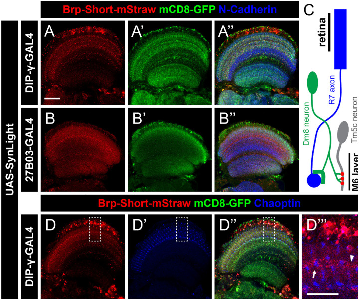
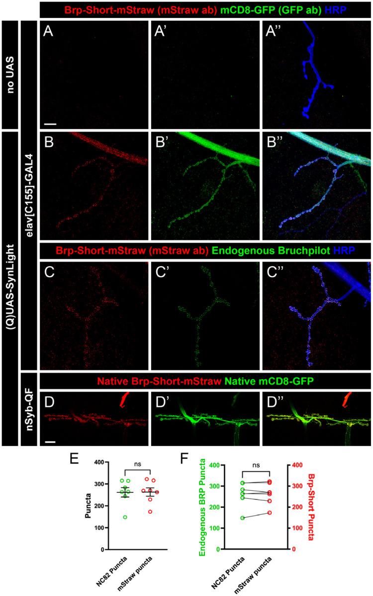
At synapses, chemical neurotransmission mediates the exchange of information between neurons, leading to complex movement behaviors and stimulus processing. The immense number and variety of neurons within the nervous system makes discerning individual neuron populations difficult, necessitating the development of advanced neuronal labeling techniques. In Drosophila , Bruchpilot-Short and mCD8-GFP, which label presynaptic active zones and neuronal membranes, respectively, have been widely used to study synapse development and organization. This labeling is often achieved via expression of two independent constructs by a single binary expression system, but expression can weaken when multiple transgenes are expressed by a single driver. Ensuring adequate expression of each transgene is essential to enable more complex experiments; as such, work has sought to circumvent these drawbacks by developing methods that encode multiple proteins from a single transcript. Self-cleaving peptides, specifically 2A peptides, have emerged as effective sequences for accomplishing this task. We leveraged 2A ribosomal skipping peptides to engineer a construct that produces both Bruchpilot-Short and mCD8-GFP from the same mRNA, which we named SynLight. Using SynLight, we visualized the putative synaptic active zones and membranes of multiple classes of olfactory, visual, and motor neurons and observed correct separation of signal, confirming that both proteins are being generated separately. Furthermore, we demonstrate proof-of-principle by quantifying synaptic puncta number and neurite volume in olfactory neurons and finding no difference between the synapse densities of neurons expressing SynLight or neurons expressing both transgenes separately. At the neuromuscular junction, we determined that synaptic puncta number labeled by SynLight was comparable to endogenous puncta labeled by antibody staining. Overall, SynLight is a versatile tool for examining synapse density in any nervous system region of interest and allows new questions to be answered about synaptic development and organization.


Conflict of interest statement

CONFLICT OF INTEREST

The authors declare no conflicts of interest.

Figures

Figure 1.
Figure 1.. Strategy for generating SynLight, a single transgene that expresses both membrane-tagged GFP and mStrawberry-tagged Bruchpilot-Short.
A, Diagram of an example plasmid containing a UAS vector and codon-optimized 2A peptide coding sequence (Daniels et al., 2014). Flanking either side of 2A are multiple cloning sites and restriction sites that facilitate insertion of two or more genes of interest. B, Diagram of the SynLight plasmid. Using restriction enzymes, the mCD8-GFP coding sequence was inserted preceding the P2A coding sequence and then the Bruchpilot-Short-mStrawberry coding sequence is inserted following the P2A sequence, keeping all sequences in frame. C, Diagram of SynLight mRNA, showing two separate proteins being produced from a single mRNA sequence. D-D”, Representative maximum projection confocal image stacks of multiglomerular LNs of the adult brain expressing SynLight and stained with antibodies against mStraw (red), GFP (green), and N-Cadherin (blue). These images show overlapping, yet distinctly different subcellular localization of Brp-Short-mStraw and mCD8-GFP. E-E”, Representative maximum projection confocal image of the third instar ventral nerve cord expressing SynLight, showing separate endogenous expression of Brp-Short-mStraw and mCD8-GFP via the native fluorescence from the mStrawberry and GFP fluorophores. Scale bars = 40 μm (D); 80 μm (E).
Figure 2.
Figure 2.. SynLight expression does not affect synapse number in olfactory neurons.
A, Diagram of the Drosophila antennal lobes showing ORNs (green) of the VA1lm glomerulus (orange). B-B”, Representative confocal image stacks of 10-day old male adult VA1lm ORNs expressing Brp-Short-mStraw and membrane-tagged GFP separately and stained with antibodies against mStraw (red), GFP (green), and N-Cadherin (blue). C-C”, High-magnification, single optical image section of ORNs from inset in B showing co-localization, but not complete overlap, of synaptic labels. D-D”, Representative confocal image stacks of 10-day old male adult VA1lm ORNs expressing SynLight and stained with antibodies as in B. E-E”, High-magnification, single optical image section from inset in D also showing co-localization, but not complete overlap, consistent with subcellular localization and suggesting P2A-mediated cleavage is occurring successfully. F-H, Quantification of Brp-Short-mStraw puncta number (F), membrane GFP volume (G), and synapse density (H) for adult male VA1lm ORNs expressing either SynLight or Brp-Short-mStraw and membrane-tagged GFP separately. Brp-Short puncta number, neurite volume, and synapse density obtained using SynLight are not significantly different from using Brp-Short-mStraw and membrane-GFP separately. For each genotype, n ≥ 20 glomeruli from 10 brains. n.s. = not significant. Scale bars = 5 μm.
Figure 3.
Figure 3.. SynLight labels presynaptic active zones and neuronal membranes in multiple cell types of the olfactory system.
A, Diagram of the Drosophila antennal lobes showing ORNs (green), PNs (magenta), and multiglomerular LNs (blue) of the DA1 glomerulus (orange). B-C”, Representative confocal image maximum projections of male adult DA1 ORNs expressing SynLight via a GAL4 (B) or QF (C) driver and stained with antibodies against mStraw (red), GFP (green), and N-Cadherin (blue). D-E”, Representative confocal image maximum projections of male adult PNs (D) and multiglomerular LNs (E) of the DA1 glomerulus expressing SynLight and stained with antibodies as in B-C. Scale bar = 5 μm.
Figure 4.
Figure 4.. SynLight labels presynaptic active zones and neuronal membranes in neurons of the visual system.
A-B”, Representative single confocal image sections of male adult brains expressing SynLight using DIP-γ-GAL4 to label Dm8 neurons (A) or 27B03-GAL4 to label optic lobe neurons (B) and stained with antibodies against mStraw (red), GFP (green), and N-Cadherin (blue). C, Schematic showing the connections between R7 photoreceptor axons (blue), Dm8 neurons (green), and Tm5c neurons (gray). R7 axons project from the retina and synapse onto the dendrites of Dm8 neurons. Dm8 neurons subsequently form synapses with Tm5c neurons, forwarding the visual information received from R7 axons. The presynaptic active zones of Dm8 neurons (red) and axon terminals of R7 cells (blue) are both found in the M6 layer of the medulla. D-D”, Representative single confocal image sections of male adult brains expressing SynLight in Dm8 neurons and stained with antibodies against mStraw (red), GFP (green), and Chaoptin (blue). D”’, Single, high-magnification image section from insets (dashed boxes, D) showing mStraw and Chaoptin co-staining. Arrow indicates region of Brp-Short and Chaoptin in close proximity while arrowhead indicates a region with only Brp-Short. Scale bars = 20 μm (A); 10 μm (D”’).
Figure 5.
Figure 5.. SynLight labels the larval neuromuscular junction and does not alter synapse formation.
A-B”, Representative confocal image maximum projections of muscle 4 NMJs in control (A) or SynLight-expressing (B) wandering third instar larvae stained with antibodies against mStraw (red), GFP (green), and HRP (blue). The negative control lacking SynLight shows no mStraw or GFP immunoreactivity while pan-neuronal SynLight expression shows clear visibility of both markers. C-C”, Representative confocal image maximum projections of a muscle 4 NMJ expressing pan-neuronal SynLight and stained for antibodies against mStraw (red), NC82 (green), and HRP (blue). D-D”, Representative confocal image maximum projections of muscle 6/7 NMJs expressing SynLight showing endogenous expression of Brp-Short-mStraw (red) and mCD8-GFP (green). E, Quantification of active zone puncta visualized by antibody staining of endogenous Bruchpilot (via monoclonal antibody NC82) or expression of Brp-Short via SynLight from C. There is no significant difference between Brp-Short-positive and NC82-positive puncta. F, Quantification of active zone puncta from C when separated into each individual NMJ corroborates there is no significant difference between Brp-Short-positive and NC82-positive puncta number. For each experimental group, n ≥ 7 NMJs. n.s. = not significant. Scale bars = 15 μm (A); 20 μm (D).

References

    1. Aimino M. A., DePew A. T., Restrepo L., & Mosca T. J. (2023). Synaptic Development in Diverse Olfactory Neuron Classes Uses Distinct Temporal and Activity-Related Programs. Journal of Neuroscience, 43(1), 28–55. 10.1523/JNEUROSCI.0884-22.2022 - DOI - PMC - PubMed
    1. Andlauer T. F. M., Scholz-Kornehl S., Tian R., Kirchner M., Babikir H. A., Depner H., Loll B., Quentin C., Gupta V. K., Holt M. G., Dipt S., Cressy M., Wahl M. C., Fiala A., Selbach M., Schwärzel M., & Sigrist S. J. (2014). Drep-2 is a novel synaptic protein important for learning and memory. ELife, 3(November), 1–24. 10.7554/eLife.03895 - DOI - PMC - PubMed
    1. Araki T., Osaka J., Kato Y., Shimozono M., Kawamura H., Iwanaga R., Hakeda-Suzuki S., & Suzuki T. (2020). Systematic identification of genes regulating synaptic remodeling in the Drosophila visual system. Genes and Genetic Systems, 95(3), 101–110. 10.1266/ggs.19-00066 - DOI - PubMed
    1. Bennett M. R. (2011). Schizophrenia: Susceptibility genes, dendritic-spine pathology and gray matter loss. Progress in Neurobiology, 95(3), 275–300. 10.1016/j.pneurobio.2011.08.003 - DOI - PubMed
    1. Berger-Müller S., Sugie A., Takahashi F., Tavosanis G., Hakeda-Suzuki S., & Suzuki T. (2013). Assessing the role of cell-surface molecules in central synaptogenesis in the Drosophila visual system. PLoS ONE, 8(12), 1–14. 10.1371/journal.pone.0083732 - DOI - PMC - PubMed

Publication types