[Preprint]. 2023 Jul 19:2023.07.18.549562.
doi: 10.1101/2023.07.18.549562.

ELASTIC FIBERS DEFINE EMBRYONIC TISSUE STIFFNESS TO ENABLE BUCKLING MORPHOGENESIS OF THE SMALL INTESTINE

Affiliations

ELASTIC FIBERS DEFINE EMBRYONIC TISSUE STIFFNESS TO ENABLE BUCKLING MORPHOGENESIS OF THE SMALL INTESTINE

Elise A Loffet et al. bioRxiv. .

Update in

Abstract

During embryonic development, tissues must possess precise material properties to ensure that cell-generated forces give rise to the stereotyped morphologies of developing organs. However, the question of how material properties are established and regulated during development remains understudied. Here, we aim to address these broader questions through the study of intestinal looping, a process by which the initially straight intestinal tube buckles into loops, permitting ordered packing within the body cavity. Looping results from elongation of the tube against the constraint of an attached tissue, the dorsal mesentery, which is elastically stretched by the elongating tube to nearly triple its length. This elastic energy storage allows the mesentery to provide stable compressive forces that ultimately buckle the tube into loops. Beginning with a transcriptomic analysis of the mesentery, we identified widespread upregulation of extracellular matrix related genes during looping, including genes related to elastic fiber deposition. Combining molecular and mechanical analyses, we conclude that elastin confers tensile stiffness to the mesentery, enabling its mechanical role in organizing the developing small intestine. These results shed light on the role of elastin as a driver of morphogenesis that extends beyond its more established role in resisting cyclic deformation in adult tissues.

Keywords: buckling; elastin; gut looping; mechanobiology; soft tissue mechanics.


Conflict of interest statement

DECLARATION OF COMPETING INTERESTS: The authors have no competing interests to disclose.

Figures

Figure 1 -
Figure 1 -. Overview of bulk RNA sequencing analysis of the dorsal mesentery during buckling morphogenesis of intestinal loops.
a. Intestines from chick embryos at E8, E12 and E16, reflecting the beginning, middle, and end, respectively, of buckling morphogenesis of intestinal loops (scale bar = 1mm); below, a schematic overview of the bulk RNAseq experiment, b. Graphic representation of buckling of the intestinal tube driven by differential growth between the tube and the mesentery; separated lengths of the tube and mesentery are shown below the intact configurations to reflect accentuation of differential elongation rates over time. This growth differential drives buckling of the intestine into loops. c. Principal Component Analysis on 1000 genes of bulk RNAseq data of mesentery tissues at E8, E12 and E16.
Figure 2 -
Figure 2 -. Bulk RNAseq reveals broad transcriptome changes related to extracellular matrix during looping.
a. Bar plot of 15 Gene Ontologies with highest E12-associated z-score for E8 vs E12 differential gene expression analysis. Z-score is calculated based on how many genes in the gene ontology are up-regulated at E12 vs E8. Arrows indicate ontologies linked to the extracellular matrix. b. Heatmap of normalized expression of genes in “extracellular matrix” gene ontology that are differentially expressed between developmental stages. c. Box plots of normalized read counts for Eln, Fbln5, Loxl2.
Figure 3 -
Figure 3 -. Elastin protein is enriched in the mesentery during looping and expression levels correlate with tissue stiffness.
a. Immunofluorescence staining for elastin and cell nuclei (DAPI, blue) on E8, E12 and E16 sections (scale bar = 100 μm). White arrowhead indicates dorsal mesentery. b. Representative stress-strain curves from uniaxial tensile testing of mesenteries at E8, E12, and E16. c-e. Toe modulus (Etoe, c), transition strain (ε*, d) and linear modulus (Elin, e) for mesenteries calculated from uniaxial tensile tests of mesentery between E8 and E16; for Elin, p < 0.0001 for E8 vs E16, p< 0.05 for all other comparisons except E8 vs E10 and E14 v E16). f. qPCR for Eln in the mesentery between E8 and E16 (p < 0.005 for E8 vs E16 and E10 vs E16, p < 0.05 for E8 vs E14, E10 vs E14, E12 vs E16). g. Correlation between expression levels of Eln and Elin, from E8 to E16 (R2 =0.5752). ns = not significant (p>0.05); *p <0.05; **p <0.005; ***p <0.0005
Figure 4 -
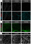
Figure 4 -. Elastase treatment degrades elastin while preserving cell viability and collagen organization in the E16 mesentery.
a. Whole-mount immunofluorescence staining for elastin and DAPI in the mesentery following incubation of intestine and mesentery segments with 0, 2, or 4 U/mL elastase (scale bar = 50 μm) b. Live/dead staining to assess cell viability, where live cells stain positively for calcein (green) and dead cells stain for ethidium homodimer 1 (EthD1, scale bar = 100 μm) c. Second Harmonic Generation (SHG) imaging of collagen network in control and elastase-treated mesenteries (scale bar = 50 μm).
Figure 5 -
Figure 5 -. Elastin depletion reduces toe and linear moduli of the mesentery.
a-c. Toe modulus (Etoe, a), transition strain (ε*, b), and linear modulus (Elin, c) for control and elastase-treated mesenteries, d. Poisson’s ratio for control and elastase-treated mesenteries measured while stress/strain curve was in the toe region, and in the linear region, n.s. = not significant (p>0.05); *p<0.05; **p<0.005; ***p<0.0005.

Similar articles

Cited by

References

    1. Goodwin K., Nelson C.M., Mechanics of Development, Dev. Cell. 56 (2021) 240–250. 10.1016/j.devcel.2020.11.025. - DOI - PMC - PubMed
    1. Collinet C., Lecuit T., Programmed and self-organized flow of information during morphogenesis, Nat. Rev. Mol. Cell Biol. 22 (2021) 245–265. 10.1038/S41580-020-00318-6. - DOI - PubMed
    1. Loffet E.A., Durel J.F., Nerurkar N.L., Evo-Devo Mechanobiology: The Missing Link, Integr. Comp. Biol. (2023) icad033. 10.1093/icb/icad033. - DOI - PubMed
    1. Heer N.C., Martin A.C., Tension, contraction and tissue morphogenesis, Development. 144 (2017) 4249–4260. 10.1242/dev.151282. - DOI - PMC - PubMed
    1. Papusheva E., Heisenberg C.-P., Spatial organization of adhesion: force-dependent regulation and function in tissue morphogenesis., EMBO J. 29 (2010) 2753–2768. 10.1038/emboj.2010.182. - DOI - PMC - PubMed

Publication types