[Preprint]. 2023 Jul 15:2023.07.13.548265.
doi: 10.1101/2023.07.13.548265.

Sustaining wakefulness: Brainstem connectivity in human consciousness

Affiliations

Sustaining wakefulness: Brainstem connectivity in human consciousness

Brian L Edlow et al. bioRxiv. .

Abstract

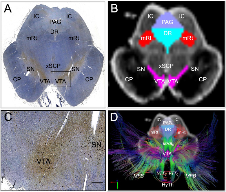
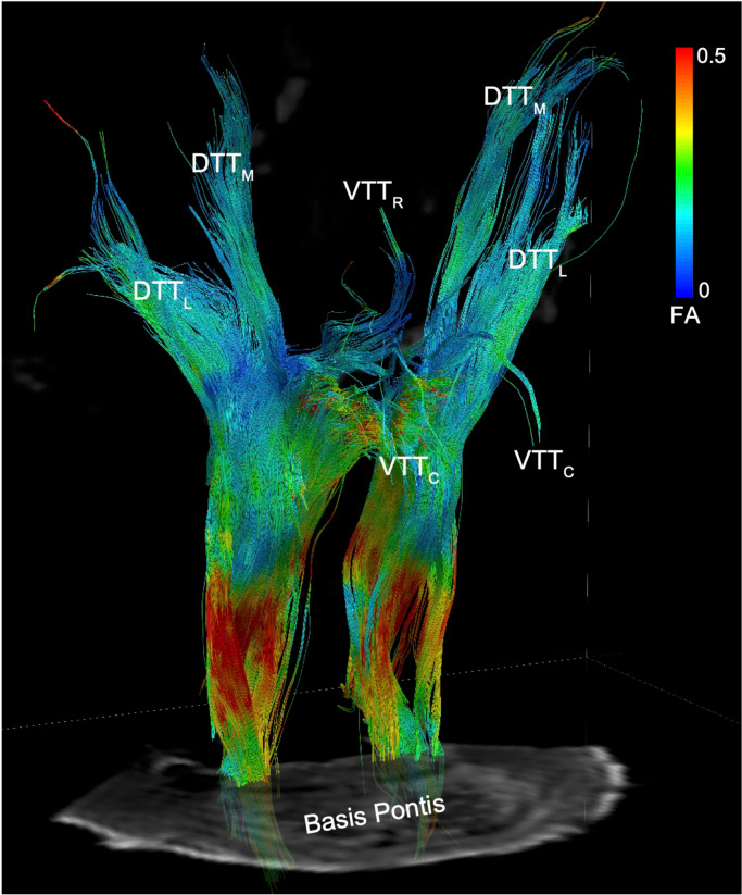
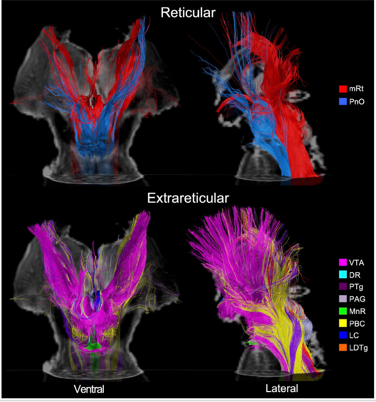
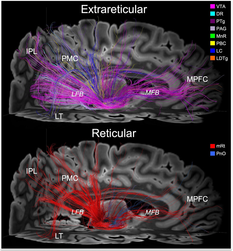
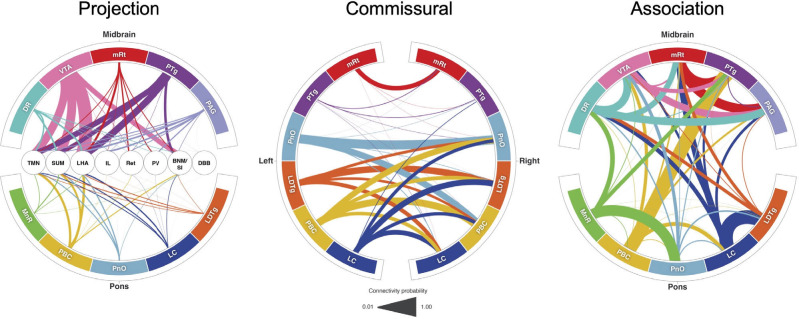
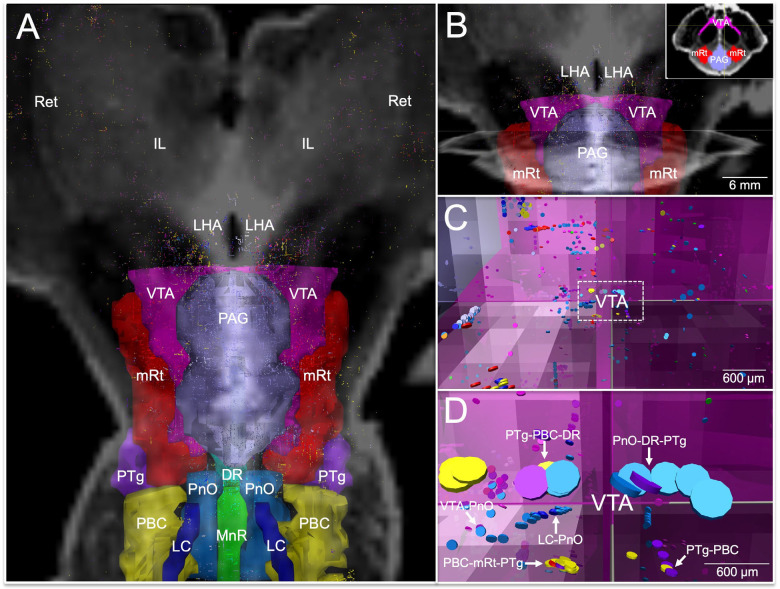
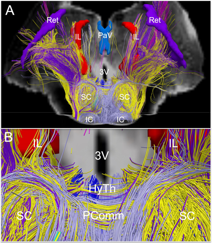
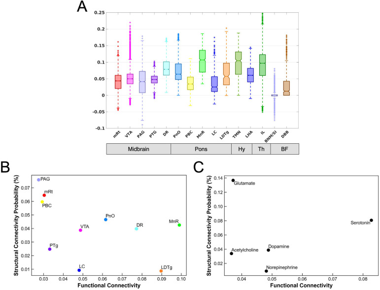
Consciousness is comprised of arousal (i.e., wakefulness) and awareness. Substantial progress has been made in mapping the cortical networks that modulate awareness in the human brain, but knowledge about the subcortical networks that sustain arousal is lacking. We integrated data from ex vivo diffusion MRI, immunohistochemistry, and in vivo 7 Tesla functional MRI to map the connectivity of a subcortical arousal network that we postulate sustains wakefulness in the resting, conscious human brain, analogous to the cortical default mode network (DMN) that is believed to sustain self-awareness. We identified nodes of the proposed default ascending arousal network (dAAN) in the brainstem, hypothalamus, thalamus, and basal forebrain by correlating ex vivo diffusion MRI with immunohistochemistry in three human brain specimens from neurologically normal individuals scanned at 600-750 μm resolution. We performed deterministic and probabilistic tractography analyses of the diffusion MRI data to map dAAN intra-network connections and dAAN-DMN internetwork connections. Using a newly developed network-based autopsy of the human brain that integrates ex vivo MRI and histopathology, we identified projection, association, and commissural pathways linking dAAN nodes with one another and with cortical DMN nodes, providing a structural architecture for the integration of arousal and awareness in human consciousness. We release the ex vivo diffusion MRI data, corresponding immunohistochemistry data, network-based autopsy methods, and a new brainstem dAAN atlas to support efforts to map the connectivity of human consciousness.


Conflict of interest statement

Competing interests: BF has a financial interest in CorticoMetrics, a company whose medical pursuits focus on brain imaging and measurement technologies. BF’s interests were reviewed and are managed by Massachusetts General Hospital and Mass General Brigham HealthCare in accordance with their conflict-of-interest policies.

Figures

Fig. 1.
Fig. 1.. Histological guidance of node localization and tract construction.
(A) A transverse section through the caudal midbrain of specimen 1 is shown, stained with tyrosine hydroxylase and counterstained with hematoxylin. The tyrosine hydroxylase stain identifies dopaminergic neurons of the ventral tegmental area (VTA). A zoomed view of VTA neurons located within the rectangle in (A) are shown in (C; scale bar = 100 μm). The corresponding non-diffusion-weighted (b=0) axial image from the same specimen is shown in (B). VTA neurons are manually traced in pink, based upon the tyrosine hydroxylase staining results, and additional arousal nuclei are traced based on the hematoxylin and eosin/Luxol fast blue stain results: DR = dorsal raphe; mRt = mesencephalic reticular formation; PAG = periaqueductal grey. Deterministic fiber tracts emanating from the VTA node are shown in (D) from the same superior view as panel (B). The tracts are color-coded by direction (inset, bottom left) and the DR and mRt nodes are semi-transparent so that VTA tracts can be seen passing through them via the mesencephalic homeostatic bundle, rostral division (MHBR). VTA tracts also connect with the hypothalamus (HyTh), thalamus, basal forebrain, and cerebral cortex via multiple bundles: MFB = medial forebrain bundle; VTTC = ventral tegmental tract, caudal division. Additional abbreviations: CP = cerebral peduncle; IC = inferior colliculus; SN = substantia nigra; xSCP = decussation of the superior cerebellar peduncles.
Fig. 2.
Fig. 2.. Reticular pathways of the human default ascending arousal network.
Deterministic tractography results from a seed region in the mesencephalic reticular formation (mRt), the brainstem region stimulated by Moruzzi and Magoun in their seminal investigations of the reticular activating system (13), are shown from a right ventro-lateral perspective in Specimen 1. Tracts are color-coded by the fractional anisotropy (FA) along each segment (inset color bar). Tracts are superimposed upon a non-diffusion-weighted (b=0) axial image at the level of the mid-pons. The fiber tracts emanating from mRt travel in the ponto-mesencephalic tegmentum and connect with the thalamus, hypothalamus, and basal forebrain via the following bundles: DTTL = dorsal tegmental tract, lateral division; DTTM = dorsal tegmental tract, medial division; VTTC = ventral tegmental tract, caudal division; VTTR = ventral tegmental tract, rostral division.
Fig. 3.
Fig. 3.. Reticular and extrareticular tracts of the default ascending arousal network.
Deterministic fiber tracts emanating from the reticular arousal nuclei are shown in the top panels. Fiber tracts emanating from extrareticular arousal nuclei are shown in the bottom panels. All tracts are from specimen 1 and superimposed on an axial non-diffusion-weighted (b=0) image at the level of the mid-pons. Additionally, the tracts in the ventral perspective (left column) are superimposed on a coronal b=0 image at the level of the mid-thalamus, and the tracts in the left lateral perspective (right column) are superimposed on a sagittal b=0 image located at the midline. All tracts are color-coded according to their nucleus of origin (inset, right). Abbreviations are described in Table 1.
Fig. 4.
Fig. 4.. Connections linking brainstem nodes of the default ascending arousal network with cortical nodes of the default mode network.
Deterministic fiber tracts emanating from the extrareticular and reticular arousal nuclei are shown in the top and bottom panels, respectively. All tracts are shown from a right lateral perspective and superimposed upon an axial non-diffusion-weighted (b=0) image at the level of the rostral midbrain and a sagittal b=0 image to the right of the midline. Tract colors and abbreviations are the same as in Fig. 3. Both the extrareticular and reticular brainstem arousal nuclei connect extensively with cortical nodes of the default mode network, including the medial prefrontal cortex (MPFC), posteromedial complex (PMC; i.e., posterior cingulate and precuneus), inferior parietal lobule (IPL), and lateral temporal lobe (LT). The primary pathway that connects the brainstem arousal nuclei to MPFC is the medial forebrain bundle (MFB). The primary pathway that connects the brainstem arousal nuclei to the PMC, IPL and LT is the lateral forebrain bundle (LFB).
Fig. 5.
Fig. 5.. Default ascending arousal network connectograms.
Projection pathways (left) connect brainstem nodes to hypothalamic, thalamic, and basal forebrain nodes. Commissural pathways (middle) connect contralateral brainstem nodes. Association pathways (right) connect ipsilateral and/or midline brainstem nodes. Brainstem dAAN nodes are shown in the outer ring of all connectograms. In the projection connectogram, the hypothalamic, thalamic and basal forebrain nodes are shown in the center: TMN = tuberomammillary nucleus; LHA = lateral hypothalamic area; IL = intralaminar nuclei; Ret = reticular nuclei; PaV = paraventricular nuclei; BNM/SI = nucleus basalis of Meynert/substantia innominata; DBB = diagonal band of Broca. Line thicknesses in each connectogram is proportional to the connectivity probability (CP) value derived from probabilistic tractography analysis. For clarity of visualization, interspecimen mean in CP measurements is shown in the connectograms. In the commissural connectogram, we show the connections from left-to-right and omit the right-to-left connections for ease of visualization. In the association connectogram, we take a mean of left- and right-sided connectivity for bilaterally represented nodes. We do not show a connectogram for projection pathways to the DMN nodes because the connectivity probability values are lower than for diencephalic and forebrain projection pathways (Table S10), such that connections would not be visualizable at this scale.
Fig. 6.
Fig. 6.. Connections linking association pathways of the default arousal network.
All candidate brainstem dAAN nodes and their tract end-points are shown from a dorsal perspective in (A), superimposed upon a coronal non-diffusion-weighted (b=0) image at the level of the mid-thalamus. Tract end-points represent the start-points and termination-points of each tract (i.e., there are two end-points per tract). All tract end-points are color-coded by the node from which they originate, and the nodes are rendered semi-transparent so that end-points can be seen within them. In (A), tract end-points are seen within each brainstem candidate node, as well as in the lateral hypothalamic area (LHA), intralaminar nuclei of the thalamus (IL), and reticular nuclei of the thalamus (Ret). A zoomed-in view of the tract end-points within the ventral tegmental area (VTA), periaqueductal grey (PAG), and mesencephalic reticular formation (mRt) is shown in (B), superimposed upon the same coronal b=0 image as in (A), as well as an axial b=0 image at the intercollicular level of the midbrain. Low-zoom and high-zoom views of tract end-points (appearing as discs) within VTA are shown in (C) and (D), respectively; the axes shown in (B, C, and D) are identical and are located at the dorsomedial border of the VTA node, as shown in the Panel B inset. In (C and D), the perspective is now ventral to the PAG, just at the dorsomedial border of the VTA, as indicated by the axes. Tract end-points from multiple candidate brainstem dAAN nodes are seen overlapping within the VTA and along its dorsal border (arrows): pedunculotegmental, parabrachial complex, and dorsal raphe (PTg-PBC-DR); pontis oralis, DR, and PTg (PnO-DR-PTg); VTA-PnO; PBC-mRt-PTg; locus coeruleus and PnO (LC-PnO); and PTg-PBC. Tract end-point overlap implies that that the beginning and/or endpoints of two tracts are in very close proximity, suggesting extensive connectivity via association pathways between ipsilateral and midline dAAN nodes with the VTA.
Fig. 7.
Fig. 7.. Extrareticular commissural and projection pathways of the default ascending arousal network.
Deterministic fiber tracts emanating from extrareticular arousal nuclei are shown from a dorsal perspective in (A) and a zoomed dorsal perspective in (B), superimposed on a coronal non-diffusion-weighted (b=0) image at the level of the mid-thalamus. Tract color-coding is identical to that in Fig. 3. Projection fibers are seen connecting with the hypothalamus (HyTh; individual hypothalamic nuclei not shown for clarity), and thalamic arousal nuclei: intralaminar nuclei (IL), paraventricular nuclei (PaV), and reticular nuclei (Ret). Commissural fibers cross the midline via the posterior commissure, with a high contribution of commissural fibers from the parabrachial complex (yellow), pedunculotegmental nucleus (dark purple), and periaqueductal grey (light purple).
Fig. 8.
Fig. 8.. Functional connectivity between subcortical nodes of the default ascending arousal network and cortical nodes of the default mode network and their relationship to structural connectivity.
(A) Boxplots of the DMN functional connectivity for each dAAN node. Y-axis represents the strength of functional connectivity (unitless) relative to the cortical DMN connectivity measured by the NASCAR method. VTA voxels showed the highest level of maximal functional connectivity with the DMN, while MnR had the highest level of median connectivity with the DMN. (B) Scatter plot between the mean of functional connectivity (x-axis) and the structural connectivity probability (y-axis) for each brainstem nucleus. (C) The counterpart grouped by the predominant type of neurotransmitter for each node: cholinergic = PTg and LDTg; dopaminergic = VTA; glutamatergic = PBC, mRt, and PnO; noradrenergic = LC; serotonergic = MnR and DR.

References

    1. Edlow B. L., Claassen J., Schiff N. D., Greer D. M., Recovery from disorders of consciousness: mechanisms, prognosis and emerging therapies. Nat Rev Neurol 17, 135–156 (2021). - PMC - PubMed
    1. Buckner R. L., DiNicola L. M., The brain’s default network: updated anatomy, physiology and evolving insights. Nat Rev Neurosci 20, 593–608 (2019). - PubMed
    1. Brown E. N., Lydic R., Schiff N. D., General anesthesia, sleep, and coma. N Engl J Med 363, 2638–2650 (2010). - PMC - PubMed
    1. Koch C., Massimini M., Boly M., Tononi G., Neural correlates of consciousness: progress and problems. Nat Rev Neurosci 17, 307–321 (2016). - PubMed
    1. Kinney H. C., Korein J., Panigrahy A., Dikkes P., Goode R., Neuropathological findings in the brain of Karen Ann Quinlan. The role of the thalamus in the persistent vegetative state. N Engl J Med 330, 1469–1475 (1994). - PubMed

Publication types