[Preprint]. 2023 Jul 13:2023.07.13.548758.
doi: 10.1101/2023.07.13.548758.

Lipid nanoparticle-mediated delivery of mRNA into the mouse and human retina and other ocular tissues

Affiliations

Lipid nanoparticle-mediated delivery of mRNA into the mouse and human retina and other ocular tissues

Cheri Z Chambers et al. bioRxiv. .

Update in

Abstract

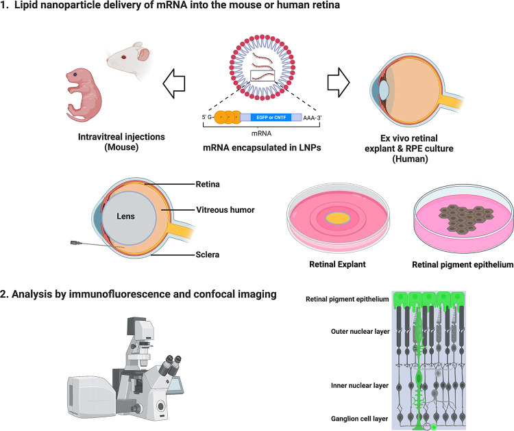
Purpose: Lipid nanoparticles (LNPs) show promise in their ability to introduce mRNA to drive protein expression in specific cell types of the mammalian eye. Here, we examined the ability of mRNA encapsulated in lipid nanoparticles (LNPs) with two distinct formulations to drive gene expression in mouse and human retina and other ocular tissues.

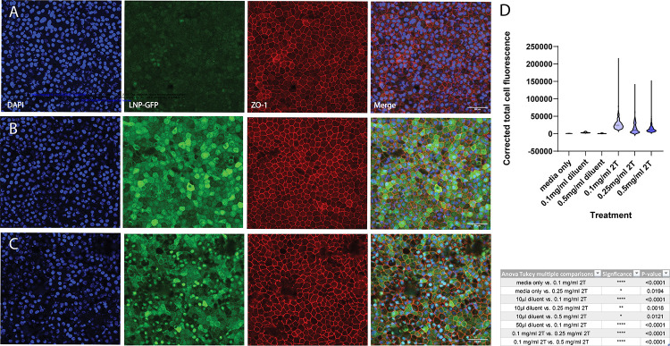
Methods: We introduced mRNA carrying LNPs into two biological systems. Intravitreal injections were tested to deliver LNPs into the mouse eye. Human retinal pigment epithelium (RPE) and retinal explants were used to assess mRNA expression in human tissue. We analyzed specificity of expression using histology, immunofluorescence, and imaging.

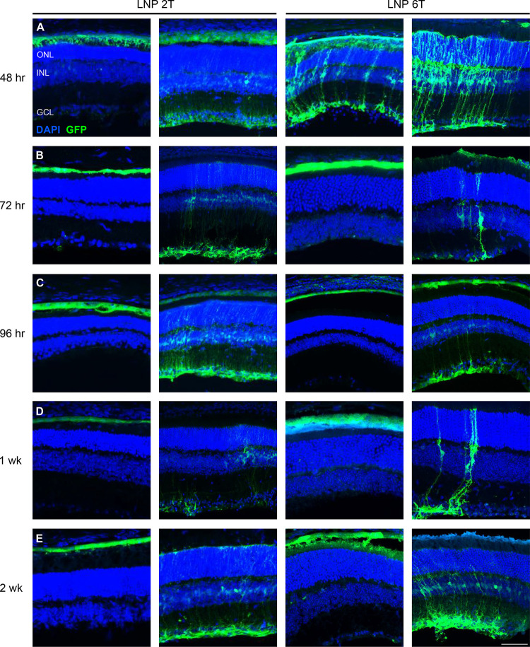
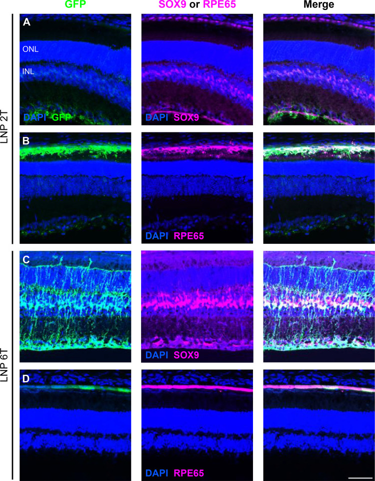
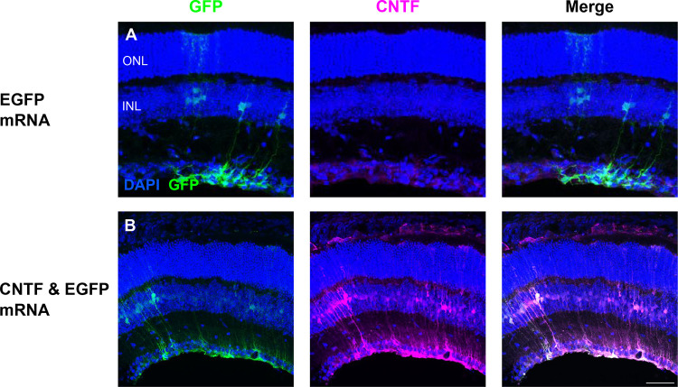
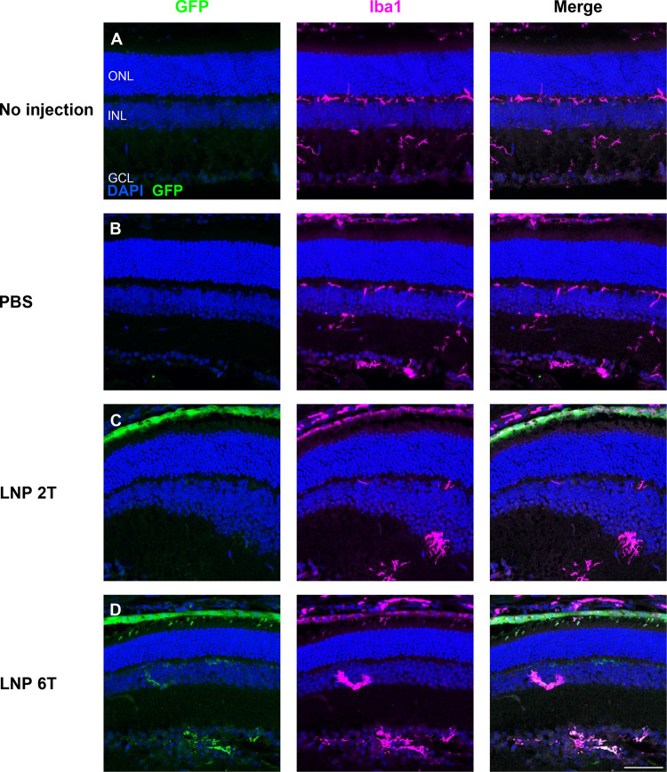
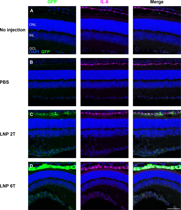
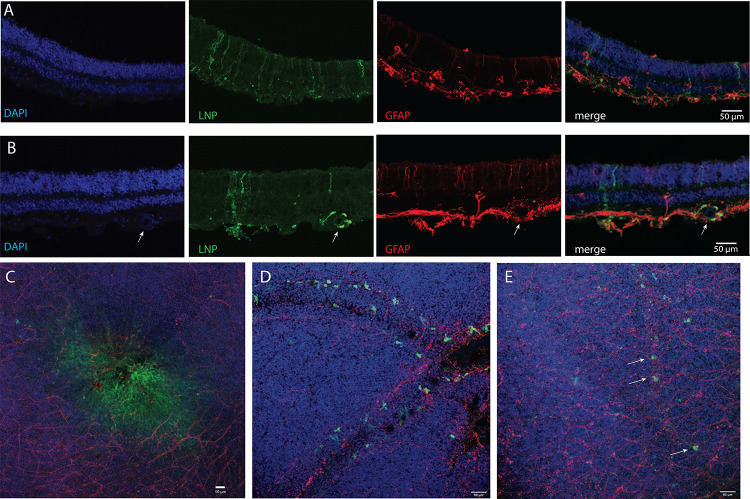
Results: In mice, mRNAs encoding GFP and ciliary neurotrophic factor (CNTF) were specifically expressed by Müller glia and retinal pigment epithelium (RPE). Acute inflammatory changes measured by microglia distribution (Iba-1) or interleukin-6 (IL-6) expression were not observed 6 hours post-injection. Human RPE also expressed high levels of GFP. Human retinal explants expressed GFP in cells with apical and basal processes consistent with Müller glia and in perivascular cells consistent with macrophages.

Conclusions: We demonstrated the ability to reliably transfect subpopulations of retinal cells in mice eye tissues in vivo and in human ocular tissues. Of significance, intravitreal injections were sufficient to transfect the RPE in mice. To our knowledge we demonstrate delivery of mRNA using LNPs in human ocular tissues for the first time.


Figures

Figure 1.
Figure 1.. Schematic describes experimental workflow to deliver mRNA encapsulated in LNPs.
CD-1 mice were used for in vivo experiments. Intravitreal injections of mRNA LNPs were performed to deliver the reagents to the vitreous chamber. After histological processing, analysis was completed using immunofluorescence and confocal imaging to determine transfection cell type expression and duration. Tile scan shows DAPI and GFP expression in a mouse retina cross-section 96 hours after intravitreal injection with EGFP mRNA encapsulated in 2TLNP. Representative image shown is a maximum intensity projection (MIP), scale bar: 500 μM.
Figure 2.
Figure 2.. Intravitreal injections of EGFP mRNA transfects retinal cells in the mouse.
Two amino lipid formulations, 2T and 6T, were compared for their ability to transfect retinal cells at various time points. Confocal images of retinal cross-sections with DAPI and EGFP staining are shown. EGFP is expressed in similar cell populations at (A) 48 hours, (B) 72 hours, (C) 96 hours, (D) 1 week, and (E) 2 weeks for both 2Tand 6T LNPs. Images shown are representative of at least 3 retinas and are MIPs (scale bar: 50 μM).
Figure 3.
Figure 3.. EGFP mRNA transfects Müller glia and RPE cells in the mouse retina.
Retinal cross-sections were co-stained with DAPI, GFP, and either SOX9 or RPE65 48 hours after intravitreal injections of EGFP mRNA encapsulated in 2Tor 6T LNPs. (A) For the 2Tformulation, nuclei labelled with EGFP overlap with nuclei labelled with SOX9, indicating EGFP expression in the Müller glia. (B) Cells that express GFP also express RPE65, indicating EGFP expression in the RPE. Similarly, for 6T, there is co-expression of EGFP and (C) SOX9 and (D) RPE65. Images shown are representative of at least 3 retinas and are MIPs (scale bar: 50 μM).
Figure 4.
Figure 4.. CNTF mRNA in 2TLNP transfects mouse Müller glia cells.
Intravitreal injections of EGFP or both EGFP and CNTF mRNA were done. (A) Control EGFP injections show EGFP expression in the Müller glia and basal CNTF expression in the GCL based on immunofluorescence staining. (B) When EGFP and CNTF mRNA were co-delivered, EGFP mRNA expression is in the Müller glia. CNTF expression is increased and in Müller glia cells as well. There is only partial overlap of EGFP and CNTF expression, indicating that there are populations of Müller glia cells that express either EGFP only, CNTF only, or both. Images shown are representative of at least 3 retinas and are MIPs (scale bar: 50 μM).
Figure 5.
Figure 5.. Immune surveillance by microglia is unchanged after intravitreal injections with EGFP mRNA LNPs.
Microglia distribution, labelled with Iba1, was compared for 4 different conditions: (A) no injection, (B) PBS intravitreal injection, (C) EGFP mRNA 2TLNP intravitreal injection, or (D) EGFP mRNA 6T LNP intravitreal injection. Microglia are present in similar patterns in the outer plexiform layer (OPL), inner plexiform layer (IPL), and GCL for all conditions 6 hours post-injection. Images shown are representative of at least 3 retinas and are MIPs (scale bar: 50 μM).
Figure 6.
Figure 6.. Intravitreal injections of EGFP mRNA LNPs does not affect the localization of cytokine IL-6.
Expression of interleukin-6 in the RPE was similar comparing 4 conditions: (A) no injection, (B) PBS controls, (C) EGFP 2TLNP, or (D) EGFP 6T LNP at 6 hours post-intravitreal injection in mouse retinas. Images shown are representative of at least 3 retinas and are MIPs (scale bar: 50 μM).
Figure 7.
Figure 7.. EGFP mRNA transfects human fRPE in vitro.
Human fRPE cells treated with EGFP mRNA encapsulated in 2TLNPs for 2 hours express EGFP. (A) fRPE cells treated with diluent only (same volume as (B)) have minimal EGFP expression. (B) Cells were treated with 0.1 mg/mL mRNA LNP and have higher GFP expression than those treated with diluent only. (C) fRPE cells treated with 0.5 mg/mL mRNA LNP also have EGFP expression higher than (A) and additionally have nuclear EGFP expression. Images shown are representative of at least 3 cell culture wells and are MIPs (scale bar: 50 μM). (D) EGFP expression was quantified and plotted in a violin plot as Corrected Total Cell Fluorescence (CTCF). P-values from an ANOVA test with post-hoc Tukey multiple comparisons are shown in the table. CTCF values are significantly higher in LNP-treated cells than in diluent-treated cells.
Figure 8.
Figure 8.. EGFP mRNA encapsulated in LNPs transfects adult human retina explants.
Adult human post-mortem eye globes were treated with EGFP mRNA LNPs for 4 hours. (A) Confocal images of human retina cross-section treated with EGFP mRNA encapsulated in 6T LNPs and then stained with DAPI, anti-GFP, and anti-GFAP. GFP is expressed in cells with apical and basal processes. (B) Cross-section of human retina treated with EGFP mRNA encapsulated in 2T LNPs. GFP is expressed in perivascular cells surrounding a blood vessel, indicated by the arrow, and in cells with apical and basal processes. (C) Flat mount of parafoveal retina treated with EGFP mRNA in 6T LNPs. GFP is expressed in cells in the fovea. (D) Flat mount of retina with RPE and choroid treated with EGFP mRNA in 2T. Perivascular cells express GFP. (E) Flat mount of retina treated with 6T. Arrows indicate GFP positive cells along a small blood vessel. Images shown are representative of retinas and RPE from two globes from a single donor (scale bars: 50 μM).

References

    1. Retinal Information Network (RetNet). https://web.sph.uth.edu/RetNet/ (2022).
    1. Hanany M., Rivolta C., Sharon D. & Daiger S. P. Worldwide carrier frequency and genetic prevalence of autosomal recessive inherited retinal diseases. doi: 10.1073/pnas.1913179117/-/DCSupplemental. - DOI - PMC - PubMed
    1. Gong J. et al. The Impact of Inherited Retinal Diseases in the US and Canada from a Cost-of-Illness Perspective. Clinical Ophthalmology 15, 2855–2866 (2021). - PMC - PubMed
    1. Fenner B. J. et al. Gene-Based Therapeutics for Inherited Retinal Diseases. Frontiers in Genetics vol. 12 Preprint at 10.3389/fgene.2021.794805 (2022). - DOI - PMC - PubMed
    1. Zhou R. & Caspi R. R. Ocular immune privilege. F1000 Biol Rep 2, 1–3 (2010). - PMC - PubMed

Publication types