[Preprint]. 2023 Jul 18:2023.07.16.549116.
doi: 10.1101/2023.07.16.549116.

Cell networks in the mouse liver during partial hepatectomy

Affiliations

Cell networks in the mouse liver during partial hepatectomy

Bin Li et al. bioRxiv. .

Update in

Abstract

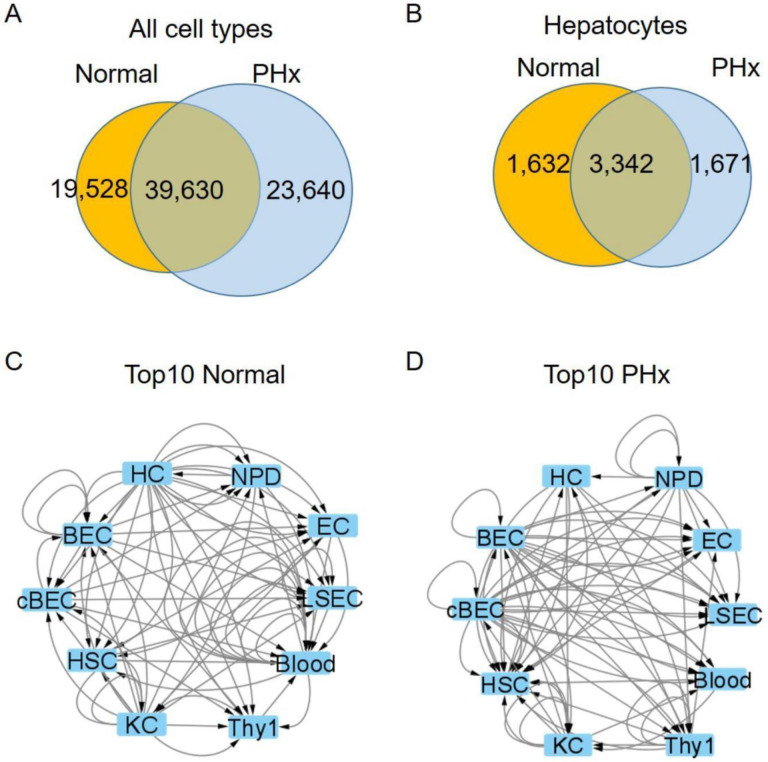
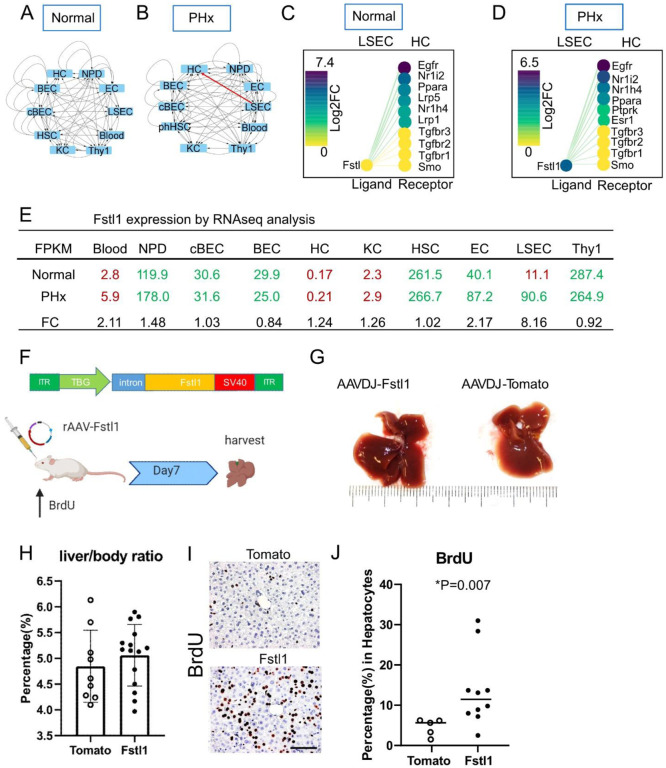
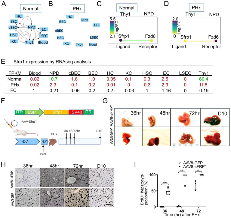
In solid tissues homeostasis and regeneration after injury involve a complex interplay between many different cell types. The mammalian liver harbors numerous epithelial and non-epithelial cells and little is known about the global signaling networks that govern their interactions. To better understand the hepatic cell network, we isolated and purified 10 different cell populations from normal and regenerative mouse livers. Their transcriptomes were analyzed by bulk RNA-seq and a computational platform was used to analyze the cell-cell and ligand-receptor interactions among the 10 populations. Over 50,000 potential cell-cell interactions were found in both the ground state and after partial hepatectomy. Importantly, about half of these differed between the two states, indicating massive changes in the cell network during regeneration. Our study provides the first comprehensive database of potential cell-cell interactions in mammalian liver cell homeostasis and regeneration. With the help of this prediction model, we identified and validated two previously unknown signaling interactions involved in accelerating and delaying liver regeneration. Overall, we provide a novel platform for investigating autocrine/paracrine pathways in tissue regeneration, which can be adapted to other complex multicellular systems.

Keywords: FACS; Fstl1; Sfrp1; Stem Cell; partial hepatectomy.


Figures

Figure 1
Figure 1. Cell-cell Interactions in homeostatic and regenerative liver
(A-B) Venn diagram of the number of paracrine and autocrine interactions in normal and partial hepatectomy (PHx) among all 10 cell types (A) and hepatocytes only (B). (A) There are 19,528 unique interactions in normal liver and 23,640 in PHx. The normal and PHx states had 39,630 shared common interactions. (B) In hepatocytes, there were 1,632 unique interactions in normal and 1,671 after PHx, with 3,342 shared common interactions. (C-D) Schematic top 10 CCInx weighed interactions in normal (C) and PHx (D) livers by Cytoscape network visualization. The interactions are ranked by the edge weights calculated by CCInx. PHx: 24 hr after 70% partial hepatectomy. HC: hepatocyte. NPD: non-progenitor duct cells. EC: Endothelial cells. LSEC: liver sinusoidal endothelial cells. Blood: blood lineage cells. Thy1: Thy1+ cells. KC: Kupffer cell. HSC: Hepatic Stellate cell. BEC: biliary epithelial cell. cBEC: clonal biliary epithelial cell. Arrows indicate either autocrine (from one cell to itself) or paracrine (from one cell to another) interactions.
Figure 2
Figure 2. Fstl1 overexpression promotes hepatocyte proliferation
(A-B) Cytoscape network visualization of Fstl1 as a ligand in 10 different cell types from the normal (A) and PHx (B) liver. Arrows indicate the paracrine and autocrine signal pathways. Red arrow indicates the paracrine signal pathway appears in the PHx but not in the normal liver. (C-D) The CCInx database showed Fstl1 as a ligand in LSEC and its top 10 receptors in HC. Fstl1 is off (yellow circle) (C) in the normal liver, but upregulated and on (dark purple circle) after partial hepatectomy (D). log2 FC: log2 fold change. (E) Normalized gene expression of Fstl1 (Tag counts in FPKM) in 10 different cell types from normal liver and PHx. Tag counts in red color: the Fstl1 gene is “off”; Tag counts in green color: the Fstl1 gene is “on”. FC: fold change. (F) rAAV construct for overexpressing the Fstl1 gene. The Fstl1 gene was cloned into a AAV2 vector backbone with the human thyroid hormone-binding globulin (TBG) as promoter. 2×10¹¹ vector genomes of AAV-Fstl1 virus were injected into C57BL/6 mice fed with BrdU in the drinking water. The liver was harvested on day 7 after injection. (G) Representative morphology of mouse liver after Fstl1 overexpression. A liver transduced with an AAVDJ-Tomato vector was used as control. (H) Liver/body weight ratio of independent mice treated with AAVDJ-Tomato (n = 9) and AAVDJ-Fstl1 (n = 15). Student’s t test. P > 0.05 (I) Histology of anti-BrdU staining in the control (Tomato) and Fstl1 overexpression liver. Scale bar = 50 μm. BrdU expressing nuclei stained brown. (J) Percentage of BrdU+ hepatocytes from Fstl1 overexpression (n = 10) and mock (Tomato) (n= 5) transduced mouse liver. Student’s t test. *P = 0.007 LSEC: liver sinusoidal endothelial cells. HC: hepatocytes
Figure 3
Figure 3. Sfrp1 delays liver regeneration
(A-B) Sfrp1 pathway in 10 different cells in the normal liver (A) and 24 hr after 70% partial hepatectomy (PHx) (B). Arrows indicate the paracrine and autocrine signal pathways. (C-D) The CCInx database showed Sfrp1 as a ligand in Thy1+ cells and its receptor Fzd6 in NPD. Log2 FC: log2 fold change. (E) Fragment per kilobase per million (FPKM) mapped reads in 10 different cell types from normal liver and PHx. Tag counts in red color: the Sfrp1 gene is “off”; Tag counts in green color: the Sfrp1 gene is “on”. FC: fold change. (F) AAV8-Sfrp1 was transfected into a wild type mouse 7 days before feeding with BrdU drinking water. One day after BrdU water treatment, partial hepatectomy was performed. Liver harvest was carried out on 36hr, 48hr, 72hr and 240hr (Day10) after partial hepatectomy. (G) Representative morphology of mouse liver overexpressing with Sfrp1 with partial hepatectomy at 36hr, 48hr, 72hr and 240hr (day10). Liver transfected with AAV8-GFP was used as control. (H) Histology of anti-BrdU staining in the mouse liver treated control (AAV8-GFP) and Sfrp1 over expressed mouse liver at different time points. Scale bar = 50 μm. BrdU expressing nuclei stained brown. (I) Quantitative assay of the percentage of Brdu+ hepatocytes in the mouse liver treated with control (AAV8-GFP, n = 3) and AAV8-Sfrp1(n = 5) at different time points. Statistical analyses: Mann-Whitney U test. **p< 0.01.

References

    1. Alaverdi N. (2004). Monoclonal antibodies to mouse cell-surface antigens. Current protocols in immunology 62, A. 4B. 1–A. 4B. 25. - PubMed
    1. Azuma H., Paulk N., Ranade A., Dorrell C., Al-Dhalimy M., Ellis E., Strom S., Kay M.A., Finegold M., and Grompe M. (2007). Robust expansion of human hepatocytes in Fah−/−/Rag2−/−/Il2rg−/− mice. Nature biotechnology 25, 903–910. - PMC - PubMed
    1. Ding B.-S., Nolan D.J., Butler J.M., James D., Babazadeh A.O., Rosenwaks Z., Mittal V., Kobayashi H., Shido K., and Lyden D. (2010). Inductive angiocrine signals from sinusoidal endothelium are required for liver regeneration. Nature 468, 310–315. - PMC - PubMed
    1. Dorrell C., Erker L., Schug J., Kopp J.L., Canaday P.S., Fox A.J., Smirnova O., Duncan A.W., Finegold M.J., and Sander M. (2011). Prospective isolation of a bipotential clonogenic liver progenitor cell in adult mice. Genes & development 25, 1193–1203. - PMC - PubMed
    1. Elzi D.J., Song M., Hakala K., Weintraub S.T., and Shiio Y. (2012). Wnt antagonist SFRP1 functions as a secreted mediator of senescence. Molecular and cellular biology 32, 4388–4399. - PMC - PubMed

Publication types