[Preprint]. 2023 Jul 20:2023.07.19.549731.
doi: 10.1101/2023.07.19.549731.

Complete Protection from SARS-CoV-2 Lung Infection in Mice Through Combined Intranasal Delivery of PIKfyve Kinase and TMPRSS2 Protease Inhibitors

Affiliations

Complete Protection from SARS-CoV-2 Lung Infection in Mice Through Combined Intranasal Delivery of PIKfyve Kinase and TMPRSS2 Protease Inhibitors

Ravi Kant et al. bioRxiv. .

Abstract

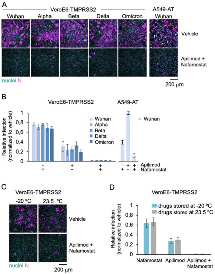
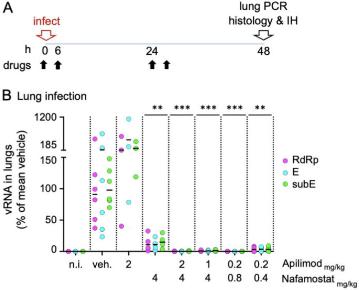
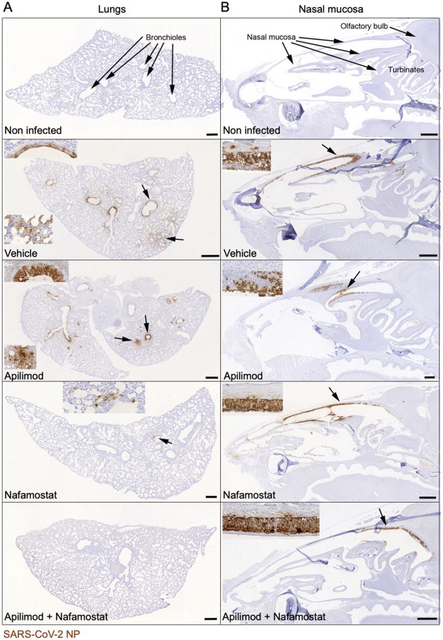
Emerging variants of concern of SARS-CoV-2 can significantly reduce the prophylactic and therapeutic efficacy of vaccines and neutralizing antibodies due to mutations in the viral genome. Targeting cell host factors required for infection provides a complementary strategy to overcome this problem since the host genome is less susceptible to variation during the life span of infection. The enzymatic activities of the endosomal PIKfyve phosphoinositide kinase and the serine protease TMPRSS2 are essential to meditate infection in two complementary viral entry pathways. Simultaneous inhibition in cultured cells of their enzymatic activities with the small molecule inhibitors apilimod dimesylate and nafamostat mesylate synergistically prevent viral entry and infection of native SARS-CoV-2 and vesicular stomatitis virus (VSV)-SARS-CoV-2 chimeras expressing the SARS-CoV-2 surface spike (S) protein and of variants of concern. We now report prophylactic prevention of lung infection in mice intranasally infected with SARS-CoV-2 beta by combined intranasal delivery of very low doses of apilimod dimesylate and nafamostat mesylate, in a formulation that is stable for over 3 months at room temperature. Administration of these drugs up to 6 hours post infection did not inhibit infection of the lungs but substantially reduced death of infected airway epithelial cells. The efficiency and simplicity of formulation of the drug combination suggests its suitability as prophylactic or therapeutic treatment against SARS-CoV-2 infection in households, point of care facilities, and under conditions where refrigeration would not be readily available.


Figures

Figure 1.
Figure 1.. The combination of apilimod dimesylate and nafamostat mesylate inhibits SARS-COV-2 and its variants in vitro, and it is stable at room temperature.
A. Representative fluorescence images of VeroE6-TMPRSS2 and A549-AT cells pre-treated with DMSO (Ctrl) or 2 μM apilimod dimesylate and 25 μM nafamostat mesylate, and one hour later infected for 20 h with indicated SARS-CoV-2 variants. Cells were stained with nuclear DNA dye Hoechst (nuclei, cyan) and immunostained with an antibody against the viral N protein (N, magenta). Scale bar = 200 μm. B-C. Quantification of the experiment shown in A. The percentage of N positive cells was determined by automated image analysis. Values represent the mean of three independent experiments and data are normalized to the infection levels obtained in DMSO vehicle treated infected cells (indicated as 1) in each experiment. The error bars represent the standard deviation. D. Representative fluorescence images of cells treated as in B with indicated drugs that had been stored either at −20 °C or room temperature (r.t.), for three months. One hour after drug treatment, cells where infected with SARS-CoV-2 Wuh strain for 20 h before fixation and immunofluorescence analysis as described in A. Scale bar 200 μm. E. Quantification of the experiment shown in D. The percentage of viral N positive cells was determined by automated image analysis. Values represent the mean of three independent experiments and data are normalized to the infection levels obtained in DMSO vehicle treated infected cells (indicated as 1) in each experiment. The error bars represent standard deviation.
Figure 2.
Figure 2.. Intranasal delivery of combined drugs at low concentrations prevents SARS-CoV-2 beta lung infection in mice.
A. Schematic description of the intranasal drug treatment and SARS-CoV-2 beta infection. Anesthetized mice received drugs intranasally in aqueous solution (30 μl) 10-20 seconds prior intranasal inoculation of SARS-CoV-2 beta (5x10⁵ plaque forming units in 20 μl DMEM). The drug treatment was repeated twice a day at 6 hours intervals at day 0 and day 1. At 48 hpi (day 2), mice were euthanized, and their right lung processed for real time quantitative PCR analysis to detect viral replication. The left lung and heads of the fixed animals were processed for immunohistology using anti N antibodies to monitor the integrity of the tissue and the distribution of viral antigens. B. PCR quantification of viral RNA in the lungs of mice treated with indicated drugs as described in A. For each mouse, the levels of viral RNA were detected using three non-overlapping primer sets, one targeting the viral genomic RNA dependent RNA polymerase gene (RdRp), and two sets against the viral gene E (E, subE). For each mouse, the obtained values were normalized first to the levels of actin in the same lung tissue and then to the mean viral RNA obtained in vehicle control treated infected mice (indicated as 100%). For each treatment, the mean (white bar) and standard deviation of the mean are indicated. The data were collected over two independent experiments each including vehicle controls. Each data point represents the RNA reads from one mouse. The concentration of apilimod dimesylate and nafamostat mesylate in mg/kg are indicated on the X axis. ***p<0.001.
Figure 3.
Figure 3.. Immunohistological analysis confirms that intranasal apilimod dimesylate nafamostat mesylate treatment prevents lung infection and limits nasal infection.
Immunohistology images of lungs and nasal mucosa from mice infected with the drugs as in Figure 2. Virus infected cells were identified with an antibody against the viral NP protein, using the horseradish peroxidase method (brown) and haematoxylin counterstain. Insets depict magnified images of the areas indicated by the arrows. Apilimod dimesylate: 2 mg/kg; nafamostat mesylate: 4 mg/kg; Apilimod dimesylate + nafamostat mesylate: 0.2 mg/kg + 0.8 mg/kg. Scale bars = 500 μm. A. Lungs. B. Nasal mucosa. Non infected mice exhibit no viral antigen in lung and nasal mucosa, whereas vehicle treated infected mice exhibit widespread SARS-CoV-2 NP expression in the lungs, both in bronchiolar epithelial cells and in pneumocytes in large groups of alveoli. This is also seen in epithelial cells in the entire nasal mucosa. In mice treated with Apilimod alone, the viral antigen expression pattern is identical, but its extent slightly reduced in the lung. After Nafamostat treatment, it is further reduced and only seen in small patches of alveolar epithelial cells. After combined Nafamostat and apilimod treatment, there is no evidence of viral antigen expression in the lung. In the nasal mucosa, positive cells are mainly seen in caudodorsal areas, in olfactory epithelial cells.
Figure 4.
Figure 4.. Intranasal delivery of combined drugs 3 h and 6 h post infection strongly decreases pulmonary cell death.
A. PCR quantification of viral RNA in the lungs of mice treated intranasally with indicated drugs at 0 h (i.e., 10-20 seconds prior infection), 3 h and 6 h post SARS-CoV-2 beta infection. For each mouse, the levels of viral RNA were detected using two primer sets, targeting the viral genomic RNA dependent RNA polymerase gene (RdRp) and the sub-genomic viral gene E (subE), respectively. For each mouse, the obtained values were normalized first to the levels of actin in the same lung tissue and then to the mean viral RNA value obtained in vehicle control-treated infected mice (indicated as 100%). For each treatment, the mean (white bar) and standard deviation of the mean are indicated. Each data point represents the RNA reads from one mouse. The concentration of apilimod dimesylate and nafamostat mesylate in mg/kg, and the time of drug administration are indicated on the X axis. B, C. Immunohistology images of lungs from mice infected and treated as described in A. Virus infected cells were identified with an antibody against the viral N protein (B, C) and apoptotic cells were visualised with an antibody against cleaved caspase 3 (C), using the horseradish peroxidase method (brown) and haematoxylin counterstain. B. Mice treated with vehicle or with nafamostat (4 mg/kg) and apilimod (2 mg/kg), starting at 3 h.p.i. or 6 h.p.i. Vehicle treated mice exhibit widespread SARS-CoV-2 NP expression in epithelial cells of bronchi (arrows) and in groups of alveoli (arrowheads). With onset of treatment at 3 h.p.i., lung infection is seen, but is less widespread than in the vehicle treated animals. With onset of treatment at 6 h.p.i., viral antigen expression is also less extensive than in the vehicle treated animals, but the reduction is less marked. Scale bars = 500 μm. C. Mice treated with vehicle or with nafamostat (4 mg/kg) and apilimod (2 mg/kg), starting at 6 h.p.i. Consecutive sections of a bronchiole stained with hematoxylin-eosin (HE), for SARS-CoV-2 NP and for cleaved caspase-3. Insets represent higher magnifications of areas indicated by the arrows in the overview images. In the vehicle treated mouse, abundant degenerate cells are present in the lumen of the bronchiole, the epithelium exhibits several degenerating cells (arrowheads in inset). There is extensive viral NP expression in epithelial cells, including degenerate cells in the lumen (arrowheads). Staining for cleaved caspase-3 shows that infected epithelial cells die via apoptosis. Inset: apoptotic epithelial cells (arrowheads); arrow: sloughed off apoptotic epithelial cell). In the nafamostat and apilimod treated animals, the bronchiolar lumina are free of degenerate cells and the epithelium appears intact, although the majority of cells are virus infected as shown by the expression of viral NP. The extreme rarity of cleaved caspase 3-positive apoptotic cells (arrow; inset: arrowhead) confirms that infected cells are viable. Scale bars = 25 μm
Figure 5.
Figure 5.. Intranasal delivery of combined drugs limits infection rebound.
A. Schematic description of the intranasal drug treatment and SARS-CoV-2 beta infection in mice. The drug treatment was repeated twice a day at 6 hours intervals at day 0 and day 1. At 48 hpi (day 2) and 96 hpi (day 4) mice were euthanized and lungs processed for PCR analysis and Immunohystochemistry to detect viral RNA and the tissue distribution of infection, respectively. B. PCR quantification of viral RNA in the lungs of mice treated intranasally with indicated drugs and infected with SARS-CoV-2 beta. For each mouse, the levels of viral RNA were detected using two primer sets, one targeting the viral genomic RNA dependent RNA polymerase (RdRp) gene and other the sub-genomic viral gene E (subE). For each mouse, the obtained values were normalized first to the levels of actin in the same lung tissue and then to the mean viral RNA value obtained in vehicle control-treated infected mice (indicated as 100%). For each treatment, the mean (white bar) and standard deviation of the mean are indicated. Each data point represents the RNA reads from one mouse. For each treatment group, the day of euthanasia is indicated on the X axis. Apilimod dimesylate 2 mg/kg, nafamostat mesylate 4 mg/kg. C. Immunohistology for SARS-CoV-2 NP in the lungs at 2 and 4 days post infection with SARS-CoV-2 beta and treated as described above. Virus infected cells were identified with an antibody against the viral N protein, using the horseradish peroxidase method (brown) and haematoxylin counterstain. Bars = 500 μm. At 2 dpi, vehicle treated mice exhibit widespread SARS-CoV-2 N expression both in bronchiolar epithelial cells and in pneumocytes in groups of alveoli. In nafamostat and apilimod treated mice, there is no evidence of viral antigen expression. At 4 dpi, the infection has been cleared in the vehicle treated mouse, i.e. there is no evidence of viral antigen expression. In mice treated with nafamostat and apilimod for the first two days after intranasal virus challenge, there are a few small groups of alveoli with viral antigen expression in pneumocytes (inset: arrowhead; the inset is a higher magnification of the area highlighted by the arrowhead in the overview image).

References

    1. Jackson C. B., Farzan M., Chen B. & Choe H. Mechanisms of SARS-CoV-2 entry into cells. Nat Rev Mol Cell Biol 23, 3–20, doi:10.1038/s41580-021-00418-x (2022). - DOI - PMC - PubMed
    1. Kreutzberger A. J. B. et al. SARS-CoV-2 requires acidic pH to infect cells. Proc Natl Acad Sci U S A 119, e2209514119, doi:10.1073/pnas.2209514119 (2022). - DOI - PMC - PubMed
    1. V'Kovski P., Kratzel A., Steiner S., Stalder H. & Thiel V. Coronavirus biology and replication: implications for SARS-CoV-2. Nat Rev Microbiol 19, 155–170, doi:10.1038/s41579-020-00468-6 (2021). - DOI - PMC - PubMed
    1. Hoffmann M. et al. SARS-CoV-2 Cell Entry Depends on ACE2 and TMPRSS2 and Is Blocked by a Clinically Proven Protease Inhibitor. Cell 181, 271–280 e278, doi:10.1016/j.cell.2020.02.052 (2020). - DOI - PMC - PubMed
    1. Koch J. et al. TMPRSS2 expression dictates the entry route used by SARS-CoV-2 to infect host cells. EMBO J 40, e107821, doi:10.15252/embj.2021107821 (2021). - DOI - PMC - PubMed

Publication types