[Preprint]. 2023 Jul 15:2023.07.14.549057.
doi: 10.1101/2023.07.14.549057.

Ebola Virus VP35 Interacts Non-Covalently with Ubiquitin Chains to Promote Viral Replication Creating New Therapeutic Opportunities

Affiliations

Ebola Virus VP35 Interacts Non-Covalently with Ubiquitin Chains to Promote Viral Replication Creating New Therapeutic Opportunities

Carlos A Rodríguez-Salazar et al. bioRxiv. .

Update in

  • Ebola virus VP35 interacts non-covalently with ubiquitin chains to promote viral replication.
    Rodríguez-Salazar CA, van Tol S, Mailhot O, Gonzalez-Orozco M, Galdino GT, Warren AN, Teruel N, Behera P, Afreen KS, Zhang L, Juelich TL, Smith JK, Zylber MI, Freiberg AN, Najmanovich RJ, Giraldo MI, Rajsbaum R. Rodríguez-Salazar CA, et al. PLoS Biol. 2024 Feb 29;22(2):e3002544. doi: 10.1371/journal.pbio.3002544. eCollection 2024 Feb. PLoS Biol. 2024. PMID: 38422166 Free PMC article.

Abstract

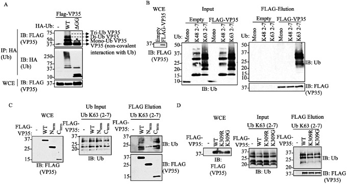
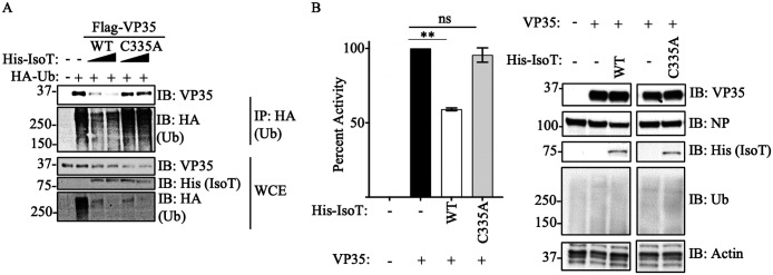
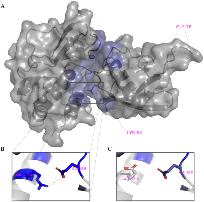
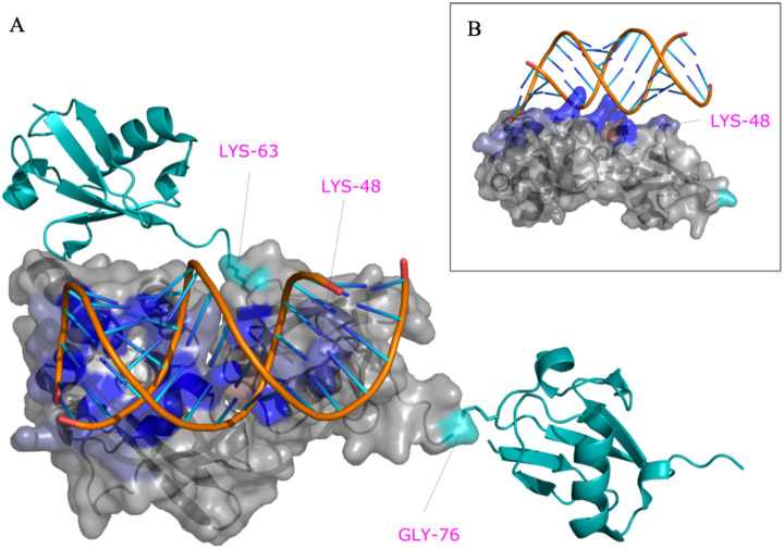
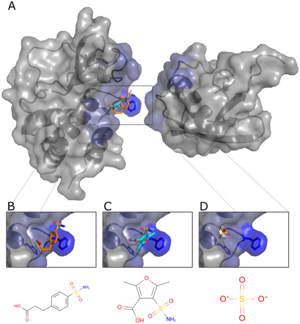
Ebolavirus (EBOV) belongs to a family of highly pathogenic viruses that cause severe hemorrhagic fever in humans. EBOV replication requires the activity of the viral polymerase complex, which includes the co-factor and Interferon antagonist VP35. We previously showed that the covalent ubiquitination of VP35 promotes virus replication by regulating interactions with the polymerase complex. In addition, VP35 can also interact non-covalently with ubiquitin (Ub); however, the function of this interaction is unknown. Here, we report that VP35 interacts with free (unanchored) K63-linked polyUb chains. Ectopic expression of Isopeptidase T (USP5), which is known to degrade unanchored polyUb chains, reduced VP35 association with Ub and correlated with diminished polymerase activity in a minigenome assay. Using computational methods, we modeled the VP35-Ub non-covalent interacting complex, identified the VP35-Ub interacting surface and tested mutations to validate the interface. Docking simulations identified chemical compounds that can block VP35-Ub interactions leading to reduced viral polymerase activity that correlated with reduced replication of infectious EBOV. In conclusion, we identified a novel role of unanchored polyUb in regulating Ebola virus polymerase function and discovered compounds that have promising anti-Ebola virus activity.

Keywords: Biological Science; Computational biology; Ebola virus; Microbiology and Biophysics and Computational Biology; Polyubiquitin; VP35; antivirals; viral polymerase.


Conflict of interest statement

Competing Interest Statement: The authors declare no competing interest.

Figures

Figure 1.
Figure 1.. EBOV VP35 protein interacts with Ub non-covalently.
A) Whole Cell extracts (WCE) from HEK293T cells transfected with Flag-VP35 (VP35) and HA-Ub wild type (WT) or HA-Ub DGG (cannot conjugate proteins), were used for HA immunoprecipitation (IP) under non-denaturing conditions (RIPA washes), followed by immunoblot (IB). B) Purified recombinant K48 or K63 polyUb chains (mix of 2–7 Ub chains), were mixed in vitro with Flag-VP35, followed by Flag IP. Interacting proteins were eluted with Flag peptide. C-D) Experiments performed as in (B) but using the C-terminal IID domain of VP35 (C), or the VP35 K309R or K309G mutants, which are not covalently ubiquitinated (D). * No ubiquitinated VP35, possibly phosphorylation.
Figure 2.
Figure 2.. Unanchored Ubiquitin interactions with VP35 promote viral polymerase activity.
A) WCE from HEK293T cells transfected with His-IsoT WT, His-IsoT C335A, VP35 WT, and HA-Ub were used for IP with anti-HA beads. B) Polymerase minigenome assay. HEK293T cells transfected with a monocistronic firefly luciferase-expressing minigenome, including VP30, L, and REN-Luc/pRL-TK, in the presence or absence of IsoT-WT or C335A mutant. Data are expressed as Mean + SEM of three independent assays in triplicate. Tukey’s multiple comparisons tests. ** p < 0.001. The percent of activity from the luciferase and renilla (Luc/ren) ratio was calculated.
Figure 3.
Figure 3.. Model of VP35 interacting with Ubiquitin.
A) The complex of VP35 (PDB ID 3JKE) and Ubiquitin (PDB ID 1UBQ) modelled using a combination of protein docking and molecular dynamics simulations. Within the complex, VP35 is shown on the left and Ubiquitin on the right. The K48 and K63 Ub residues are shown in cyan on the bottom left and C-terminal on the right within Ub. B) One of the strongest interactions contributing to the stability of the complex is ARG225-GLU18. C) Mutation of ARG225 to GLU affects interactions.
Figure 4.
Figure 4.. An intact R225 residue on VP35 is required for optimal interaction with Ub and viral polymerase function.
A) HEK293T cells were transfected with minigenome plasmids and VP35 WT, VP35 R225E, or VP35 R225K, followed by Luciferase assay. B) HEK293T cells were transfected with plasmids encoding Flag-VP35 WT, VP35 R225E, or VP35 R225K. WCE were then used to isolate Flag-tagged proteins using anti-Flag beads. After washes, the beads containing VP35 were mixed with the WCE containing HA-Ub to test binding. C) as in B, but instead of mixing with WCE, binding was performed using purified recombinant K63-linked polyUb chains, followed by Flag elution. Quantification by densitometry of 3 independent experiments is shown.
Figure 5.
Figure 5.. VP35-PolyUb-dsRNA predicted complex.
The predicted structure of the VP35-Ub complex was used as a template to superpose the structure of VP35 bound to RNA (PDB ID 3KS8). PolyUb was modeled using as a template the structure of K63 Di-Ubiquitin (PDB ID 2JF5). The residues K63, K48, and G76 of the central Ub bound to VP35 are labeled in magenta and contribute favorably to RNA binding in this model.
Figure 6.
Figure 6.. Predicted poses of small-molecules disrupting the VP35-Ub interaction.
A. The cavity within the interface with Ub with predicted bound pCEBS (B) and SFC (C) in close proximity to a sulfate ion (D) observed experimentally (PDB ID 4IBG).
Figure 7.
Figure 7.. pCEBS and SFC compounds inhibit interactions between VP35 and K63-linked polyubiquitin chains and correlate with loss of viral polymerase activity and virus replication.
A) Flag-VP35 bound to anti-Flag beads were incubated for 1h at room temperature with different concentrations of pCEBS or SFC, followed by incubation with recombinant purified unanchored K63-linked polyUb chains (–7). VP35-Ub complexes were eluted with Flag-peptide and analyzed by Immunoblot. B) 293T cells were transfected with minigenome components and 4 hours post-transfection cells were treated with pCEBS and SFC compounds at different concentrations. 50 hours later cells were lysed for luciferase assay. C) Cytotoxicity test (CyQUANT MTT Cell Viability Assay ThermoFhisher) using pCEBS and SFC at different dilutions D) Plaque reduction and (E) Virus Yield Reduction assays, the cells were infected by 1 hour and after 1h the treatment was made with pcEBS, SFB compound or DMSO: Dimethyl sulfoxide with the overlay. The number of plaques in each set of compound dilution were converted to a percentage relative to the untreated virus control. The percent of activity from the ratio of luciferase and renilla (Luc/ren) was calculated. Data are depicted as Mean + SEM of the two independent assays in triplicate. Tukey’s multiple comparisons tests. p < 0.001 **, p < 0.0001 ***, p < 0.00001 ****.

Similar articles

References

    1. Jacob S. T., et al., Ebola virus disease. Nat Rev Dis Primers 6, 13 (2020). - PMC - PubMed
    1. Baseler L., Chertow D. S., Johnson K. M., Feldmann H., Morens D. M., The Pathogenesis of Ebola Virus Disease*. Annual Review of Pathology: Mechanisms of Disease 12, 387–418 (2017). - PubMed
    1. Malvy D., McElroy A. K., de Clerck H., Günther S., van Griensven J., Ebola virus disease. The Lancet 393, 936–948 (2019). - PubMed
    1. Bharaj P., et al., The Host E3-Ubiquitin Ligase TRIM6 Ubiquitinates the Ebola Virus VP35 Protein and Promotes Virus Replication. J Virol 91, 1–17 (2017). - PMC - PubMed
    1. Leung D. W., et al., Structure of the Ebola VP35 interferon inhibitory domain. Proceedings of the National Academy of Sciences 106, 411–416 (2009). - PMC - PubMed

Publication types