Case Reports
. 2021;15(1):38-45.
doi: 10.5797/jnet.cr.2020-0015. Epub 2020 Aug 28.

A Patient with Moyamoya Disease Who Underwent Recanalization Therapy for Acute Intracranial Internal Carotid Artery Occlusion

Affiliations
Case Reports

A Patient with Moyamoya Disease Who Underwent Recanalization Therapy for Acute Intracranial Internal Carotid Artery Occlusion

Shu Sogabe et al. J Neuroendovasc Ther. 2021.

Abstract

Objective: We report a case of acute internal carotid artery occlusion in a patient with adult-onset moyamoya disease who underwent mechanical thrombectomy and had a good outcome.

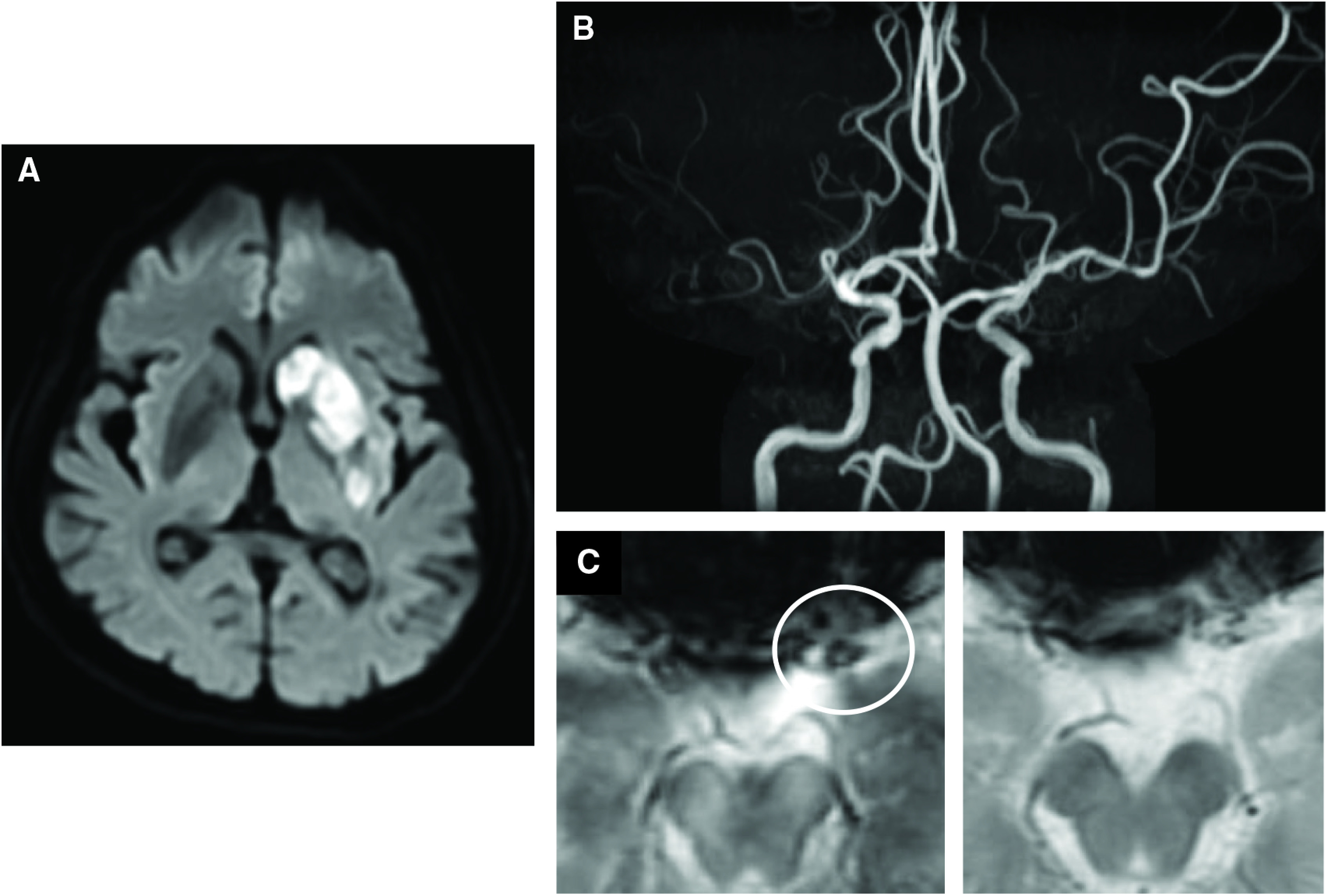
Case presentation: A 73-year-old woman was diagnosed with moyamoya disease by asymptomatic right middle cerebral artery occlusion at 59 years of age. The patient was transported for stroke symptoms. Magnetic resonance imaging (MRI) demonstrated left terminal internal carotid artery occlusion and low-intensity signal on T2*-weighted imaging at the occlusion site. Alteplase was administered and endovascular treatment was subsequently performed. A small-diameter microcatheter was guided to the distal end of the occlusion and angiography after deployment of a stent retriever revealed irregular stenosis. Severe stenosis remained after thrombectomy, and balloon angioplasty was added. The treatment resulted in recanalization and good outcome.

Conclusion: Adults with moyamoya disease may have accompanying atherosclerotic intracranial artery occlusion. Angiography after deployment of a stent retriever was useful for clarifying the etiology of occlusion. It is important to determine the etiology of occlusion based on the medical history or imaging findings and to select an appropriate treatment.

Keywords: acute recanalization therapy; moyamoya disease; percutaneous intracranial angioplasty; percutaneous intracranial thrombectomy.


Conflict of interest statement

The authors declare no conflict of interest.

Figures

Fig. 1
Fig. 1. (A) MRI DWI at the initial episode. High-intensity foci are observed in the left watershed region. (B) DSA of the right common carotid artery showing stenosis of the terminal right internal carotid artery, occlusion of the middle cerebral artery, and moyamoya vessels in the basal ganglia region. Well-developed anterior cerebral artery and collaterals are observed. (C) DSA of the left common carotid artery. Severe stenosis is observed from the terminal left internal carotid artery to the proximal middle cerebral artery. DSA: digital subtraction angiography; MRI DWI: magnetic resonance imaging diffusion-weighted imaging
Fig. 2
Fig. 2. (A) MRI DWI at the present episode. Mild high-intensity foci are observed in the left caudate nucleus and putamen. (B) MRA showing occlusion of the terminal left internal carotid artery. (C) T2*WI showing low-intensity foci considered a SVS in the terminal left internal carotid artery (white circle). MRA: magnetic resonance angiography; MRI DWI: magnetic resonance imaging diffusion-weighted imaging; SVS: susceptibility vessel sign
Fig. 3
Fig. 3. Angiography. (A) Right common carotid arteriography. The findings are nearly identical to those of previous angiography. (B) Left vertebral arteriography showing left-side-dominant development of the posterior cerebral artery and collaterals. (C) Left common carotid arteriography showing occlusion of the terminal internal carotid artery and development of the external carotid artery. (D) An enlarged view of the terminal internal carotid artery. Interruption of the blood flow and filling defect suggestive of thrombus are observed.
Fig. 4
Fig. 4. Endovascular treatment. (A) Microcatheter angiography distal to the site of occlusion. The diameter of the peripheral vessel remains nearly normal. (B) After deployment of the Tron FX. Immediate flow restoration is observed. The vascular wall at the site of occlusion is irregular. (C) After retrieval of the Tron FX. Although the internal carotid artery was recanalized, findings of severe stenosis persisted. (D) After balloon angioplasty. The site of stenosis is dilated. (E) Retrieved thrombus. The thrombus consisted of red blood cells, neutrophils, and fibrin.
Fig. 5
Fig. 5. (A) Postoperative MRI DWI. Clear high-intensity foci are observed in the basal ganglia region. (B) Postoperative MRA. Findings of recanalization of the left middle cerebral artery remain. (C) Preoperative (left) and postoperative (right) T2*WI. The low-intensity foci preoperatively observed at the occluded vessel disappeared after surgery. MRA: magnetic resonance angiography; MRI DWI: magnetic resonance imaging diffusion-weighted imaging

Similar articles

Cited by

References

    1. Tominaga T, Suzuki N, Miyamoto T, et al. . Recommendations for the management of moyamoya disease: a statement from research committee on spontaneous occlusion of the circle of Willis (Moyamoya disease) [2nd edition]. Surg Cereb Stroke 2018; 46: 1–24. (in Japanese)
    1. Suzuki J, Takaku A: Cerebrovascular “moyamoya” disease. Disease showing abnormal net-like vessels in base of brain. Arch Neurol 1969; 20: 288–299. - PubMed
    1. Hosoda Y, Ikeda E, Hirose S. Histopathological studies on spontaneous occlusion of the circle of Willis (cerebrovascular moyamoya disease). Clin Neurol Neurosurg 1997; 99 Suppl 2: S203–S208. - PubMed
    1. Liu W, Morito D, Takashima S, et al. : Identification of RNF213 as a susceptibility gene for moyamoya disease and its possible role in vascular development. PLoS ONE 2011; 6: e22542. - PMC - PubMed
    1. Lee SJ, Ahn JY: Stenosis of the proximal external carotid artery in an adult with moyamoya disease: moyamoya or atherosclerotic change? Neurol Med Chir (Tokyo) 2007; 47: 356–359. - PubMed

Publication types