Review
. 2023 Dec;39(16-18):1167-1184.
doi: 10.1089/ars.2023.0350. Epub 2023 Sep 7.

NAD+, Axonal Maintenance, and Neurological Disease

Affiliations
Review

NAD+, Axonal Maintenance, and Neurological Disease

Athanasios S Alexandris et al. Antioxid Redox Signal. 2023 Dec.

Abstract

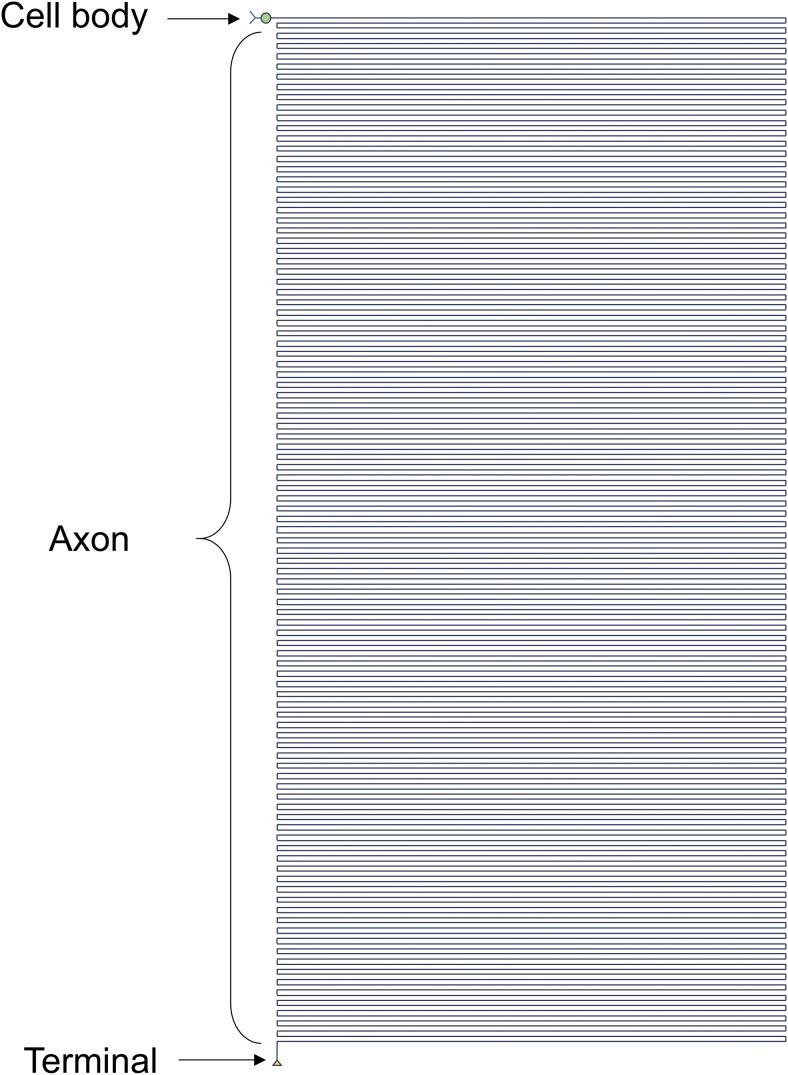
Significance: The remarkable geometry of the axon exposes it to unique challenges for survival and maintenance. Axonal degeneration is a feature of peripheral neuropathies, glaucoma, and traumatic brain injury, and an early event in neurodegenerative diseases. Since the discovery of Wallerian degeneration (WD), a molecular program that hijacks nicotinamide adenine dinucleotide (NAD+) metabolism for axonal self-destruction, the complex roles of NAD+ in axonal viability and disease have become research priority. Recent Advances: The discoveries of the protective Wallerian degeneration slow (WldS) and of sterile alpha and TIR motif containing 1 (SARM1) activation as the main instructive signal for WD have shed new light on the regulatory role of NAD+ in axonal degeneration in a growing number of neurological diseases. SARM1 has been characterized as a NAD+ hydrolase and sensor of NAD+ metabolism. The discovery of regulators of nicotinamide mononucleotide adenylyltransferase 2 (NMNAT2) proteostasis in axons, the allosteric regulation of SARM1 by NAD+ and NMN, and the existence of clinically relevant windows of action of these signals has opened new opportunities for therapeutic interventions, including SARM1 inhibitors and modulators of NAD+ metabolism. Critical Issues: Events upstream and downstream of SARM1 remain unclear. Furthermore, manipulating NAD+ metabolism, an overdetermined process crucial in cell survival, for preventing the degeneration of the injured axon may be difficult and potentially toxic. Future Directions: There is a need for clarification of the distinct roles of NAD+ metabolism in axonal maintenance as contrasted to WD. There is also a need to better understand the role of NAD+ metabolism in axonal endangerment in neuropathies, diseases of the white matter, and the early stages of neurodegenerative diseases of the central nervous system. Antioxid. Redox Signal. 39, 1167-1184.

Keywords: SARM1; Wallerian degeneration; axonal degeneration; neurodegeneration; nicotinamide; pellagra.


Conflict of interest statement

No competing financial interests exist.

Figures

FIG. 1.
FIG. 1.
The outstanding geometry of the axon. This diagram shows the relative length of the axon of a human motor neuron drawn to scale with its cell body diameter. For a similar approach, see also Devor (1999) and Koliatsos and Alexandris (2019).
FIG. 2.
FIG. 2.
Chemical structure of NAD+ and related metabolites. Niacin (red) typically refers to Na and its amide, Nam. Biosynthetic pathways can utilize either form to create amidated and nonamidated derivatives. Phosphoribosylation of niacin produces NaMN and NMN, and the further addition of an adenosine monophosphate group gives rise to NaAD and NAD. Dephosphorylation of NaMN or NMN can generate NaR and NR, respectively. For mammalian biosynthetic pathways, see Figure 3. Na, nicotinic acid; NaAD, nicotinic acid adenine dinucleotide; NAD+, nicotinamide adenine dinucleotide (oxidized); Nam, nicotinamide; NaMN, nicotinic acid mononucleotide; NaR, nicotinic acid riboside; NMN, nicotinamide mononucleotide; NR, nicotinamide riboside.
FIG. 3.
FIG. 3.
NAD+ biosynthetic pathways in mammalian cells. There are three main NAD+ biosynthetic pathways, each starting with a different NAD+ precursor. The core Nam recycling pathway (blue box) utilizes Nam produced from intracellular NAD+ degradation or imported from the extracellular space. Nam is converted to NMN by NAMPT, and this is in turn converted to NAD+ by any of the rate-limiting isoforms of the NMN adenylyltransferase enzymes (NMNAT1–3). In a manner similar to the Nam recycling pathway, the Na salvage pathway (also known as Preiss–Handler pathway; yellow box) converts Na to NaMN by NAPRT, and NaMN to NaAD by NMNAT. NaAD is then converted to NAD+ by NADSYN. In the de novo synthesis pathway (green box), tryptophan is converted to QA, which is then utilized for NAD+ synthesis via NaMN and NaAD intermediates. Extracellular NaR and NR can also be imported and utilized intracellularly after phosphorylation by NRK 1 or 2, generating NaMN or NMN, respectively (gray striped box). NAD+ exists in equilibrium with its reduced and phosphorylated forms, and can be degraded by different NAD+-consuming enzymes (see Essentials of NAD+ Metabolism). While most of intracellular Nam is recycled, it can also be eliminated and excreted after its methylation by NNMT to form MNA. MNA, 1-methylnicotinamide; NADSYN, NAD+ synthase; NAMPT, nicotinamide phosphoribosyltransferase; NAPRT, nicotinate phosphoribosyltransferase; NMNAT, nicotinamide mononucleotide adenylyltransferase; NNMT, nicotinamide n-methyltransferase; NRK, nicotinamide riboside kinase; PNP, purine nucleoside phosphorylase; QA, quinolinic acid; QPRT, quinolinate phosphoribosyl transferase.
FIG. 4.
FIG. 4.
Regulation of NAD+ pools across subcellular compartments. In axons, the core Nam recycling pathway is the main source of NAD+. NAD+ and NMN do not cross cellular membranes readily, and form distinct subcellular pools. The specifics of mitochondrial NAD+ biogenesis are debated, but NAD+ may be imported/exported via putative NAD+ transporters, while NAD+ might be also generated from imported NMN or mitochondrial Nam. Extracellular NAD+ precursors and NAD+ are further regulated by ectoenzymes such as CD 38, CD73, and CD157. While NAD+ levels are independently regulated in different subcellular compartments, NAD+ pools may affect each other via effects on NAD+ precursors or possibly via NAD+ transporters. Based on Cambronne and Kraus (2020) and Gasparrini et al. (2021). ARTC, ADP-ribosyltransferase C2/C3 toxin-like; eNAMPT, extracellular nicotinamide phosphoribosyltransferase; iNAMPT, intracellular nicotinamide phosphoribosyltransferase; NPP1, nucleotide pyrophosphatase/phosphodiesterase 1.
FIG. 5.
FIG. 5.
Axon reaction (chromatolysis) in pellagra. Nissl-stained neurons from patients with pellagrous encephalopathy are reproduced from Singer and Pollock (1913). Cells 1 and 2 are from cerebral cortex, cell 3 is from the ventral horn, and cell 4 is from a sympathetic ganglion. Axon reaction is a classical marker of axonal injury.
FIG. 6.
FIG. 6.
WD after axotomy. A transected axon of a mouse DRG neuron is shown at different time points after injury (h:mm). Primary mouse DRG neurons were obtained from YFP-16 mice and cultured in microfluidic devices for separation of cell bodies (not shown; toward the bottom of the field of view) and axons. Axotomy was performed by a razor blade and axon segments distal to the injury were visualized based on their YFP fluorescence. The distal stump shows limited retraction in the first 4.5 h (arrowheads), before a lengthwide fragmentation and subsequent dissolution at 5–6 h. DRG, dorsal root ganglion; WD, Wallerian degeneration.
FIG. 7.
FIG. 7.
A model of SARM1 activation. (A) SARM1 is a multidomain protein that forms an octamer. SARM1 has a mitochondrial localization sequence (not shown), an ARM domain (yellow) that contains sites for allosteric binding of NAD+ and NMN, two SAM domains (blue), which facilitate multimerization, and a TIR domain with NAD(P)+ glycohydrolase activity (green). The SAM domains are assembled in an inner ring, while the ARM and TIR domains radiate outward. NAD+ and NMN compete for binding at allosteric sites in the ARM domain. NAD+ binding stabilizes ARM-TIR and ARM-SAM domain interactions, and prevents TIR dimerization. NMN binding induces a conformational change that allows TIR dimerization and enzymatic activation. (B) Types of reactions catalyzed by SARM1. Based on Icso and Thompson (2022). ARM, armadillo repeat; SAM, sterile alpha motif; SARM1, sterile alpha and TIR motif containing 1; TIR, toll-interleukin-1 receptor.
FIG. 8.
FIG. 8.
WD signaling pathways. (A) Core players are SARM1 as the instructor and executor of WD and NMNAT2 as an indirect suppressor of SARM1 activity. NMNAT2 levels are regulated by a balance between axonal delivery and degradation. (B) WD signaling (red arrows) can be initiated by different types of axonal perturbations (yellow arrows) that result in loss of NMNAT2 activity. This can be achieved via direct injury to the axon and loss of anterograde axon transport, and/or increased degradation of NMNAT2 secondary to increased activity of the DLK-JNK cascade (yellow box). The DLK-JNK cascade can be activated by different abiotic and biotic stressors (yellow arrows), including direct axonal injury and probably mitochondrial impairment, and can be self-amplified. Loss of NMNAT2 activity results in accumulation of NMN, reduced NAD+ synthesis, and allosteric activation of SARM1 due to a decrease in the NAD+/NMN ratio. SARM1 activation results in local NAD+ degradation in a feed-forward manner and generation of Ca2+ mobilizers. JNK-mediated SARM1 phosphorylation may further increase its activity. Severe NAD+ depletion may also lead to mitochondrial depolarization and bioenergetic failure, and loss of both cytosolic and mitochondrial ATP synthesis; as a consequence, intracellular Ca2+ homeostasis cannot be maintained, resulting in calpain activation, proteolysis of axonal proteins, and axonal degeneration. DLK, dual leucine zipper kinase; JNK, c-JUN N-terminal kinase.
FIG. 9.
FIG. 9.
Models of axonal injury and degeneration related to WD signaling. (A) A healthy axon is subjected to an acute or subacute axonal insult (lightening strike). The axon is natively resistant to minor insults and the transient turn on of WD signaling, principally activation of SARM1 (line 1), but more severe insults that drive SARM1 activity to critical levels (lines 2 and 3) trigger feed-forward cascades and eventually axon degeneration (also see Fig. 8). Inhibition of SARM1 within a critical window may abort progression of WD and allow the axon to recover (line 2). (B) The threshold that triggers WD depends on several factors including the availability and turnover of NMNAT. Change in threshold, for example, due to transient or chronic deficits in NMNAT2 activity or NAD+ metabolism (arrows) may predispose axons to WD with future mild insults or small variations in SARM1 activity.

Similar articles

Cited by

References

    1. Adalbert R, Coleman MP. Review: Axon pathology in age-related neurodegenerative disorders. Neuropathol Appl Neurobiol 2013;39, 90–108. - PubMed
    1. Adalbert R, Nogradi A, Szabo A, et al. . The slow Wallerian degeneration gene in vivo protects motor axons but not their cell bodies after avulsion and neonatal axotomy. Eur J Neurosci 2006;24:2163–2168. - PubMed
    1. Agledal L, Niere M, Ziegler M. The phosphate makes a difference: Cellular functions of NADP. Redox Rep 2010;15:2–10. - PMC - PubMed
    1. Ahn BH, Kim HS, Song S, et al. . A role for the mitochondrial deacetylase Sirt3 in regulating energy homeostasis. Proc Natl Acad Sci U S A 2008;105:14447–14452. - PMC - PubMed
    1. Aksoy P, White TA, Thompson M, et al. . Regulation of intracellular levels of NAD: A novel role for CD38. Biochem Biophys Res Commun 2006;345:1386–1392. - PubMed

Publication types

MeSH terms