Review
. 2023 Jun 28;13(7):688.
doi: 10.3390/bios13070688.

Progress in Isolation and Molecular Profiling of Small Extracellular Vesicles via Bead-Assisted Platforms

Affiliations
Review

Progress in Isolation and Molecular Profiling of Small Extracellular Vesicles via Bead-Assisted Platforms

Daria Kozhevnikova et al. Biosensors (Basel). .

Abstract

Tremendous interest in research of small extracellular vesicles (sEVs) is driven by the participation of vesicles in a number of biological processes in the human body. Being released by almost all cells of the body, sEVs present in complex bodily fluids form the so-called intercellular communication network. The isolation and profiling of individual fractions of sEVs secreted by pathological cells are significant in revealing their physiological functions and clinical importance. Traditional methods for isolation and purification of sEVs from bodily fluids are facing a number of challenges, such as low yield, presence of contaminants, long-term operation and high costs, which restrict their routine practical applications. Methods providing a high yield of sEVs with a low content of impurities are actively developing. Bead-assisted platforms are very effective for trapping sEVs with high recovery yield and sufficient purity for further molecular profiling. Here, we review recent advances in the enrichment of sEVs via bead-assisted platforms emphasizing the type of binding sEVs to the bead surface, sort of capture and target ligands and isolation performance. Further, we discuss integration-based technologies for the capture and detection of sEVs as well as future research directions in this field.

Keywords: beads; exosomes; isolation; liquid biopsy; molecular characterization; small extracellular vesicles.


Conflict of interest statement

The authors declare no conflict of interest.

Figures
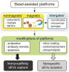
Figure 1
Figure 1
Classification of bead-assisted platforms based on nonmagnetic particles, magnetically responsive beads and those integrated with microfluidics, microarrays and porous materials. Middle row depicts the functionalization of bead-assisted platforms by coatings and organic molecules, leading to nonspecific and immunoaffinity enrichment of sEVs.
Figure 2
Figure 2
Workflow of isolation and purification of small extracellular vesicles using heparin-coated agarose beads. (i) The beads are incubated with sEVs derived from different cell lines, (ii) formation of a sEVs/bead complex, (iii) removing proteins and nucleic acids by washing with PBS, (iv) release of sEVs from the beads using a concentrated salt solution and (v) extraction of RNA from sEVs for analysis. Reprinted with permissions from reference [29]. Copyright 2015, Springer Nature.
Figure 3
Figure 3
Workflow of two-step magnetic bead-based (2MBB) method for isolation of a subset of sEVs and sEV microRNAs. Anti-CD63 antibody-coated magnetic beads are first added to human plasma samples to capture sEVs. The isolated sEVs are then eluted for size and concentration analysis and lysed for analyzing proteins and RNAs. miRNAs are extracted using oligonucleotide-conjugated magnetic beads (upper panel). Levels of miRNAs hsa-miR-21-5p (left) and hsa-miR-126-3p (right) extracted from platelet-poor plasma, sEVs captured on magnetic beads and supernatant after magnetic beads relative to the spike-in exogenous cel-miR-238-3p (bottom panel). (** p < 0.01, * p < 0.05, Student’s t test). Reprinted with permission from reference [33]. Copyright 2020, PLOS ONE.
Figure 4
Figure 4
Schematic of the fabrication of Nano pom-poms capture platform for trapping of sEV and their release via on-demand photo-cleavage (a). TEM and SEM images of Nano pom-poms and a commercial nanoplatform (b). TEM image of captured sEVs on the surface of Nano pom-poms. Red arrows indicate captured sEVs with bound 10 nm antiCD63 gold nanoparticles (c). Nanoparticle tracking analysis of sEVs isolated by Nano pom-poms and ultracentrifugation (d). Nanoparticle tracking analysis of the size of sEVs isolated with Nano pom-poms and ExoEasy kit (e). SEM images of Nano pom-poms with captured sEVs and those after the release of sEVs via on-demand photo-cleavage. Workflow of multi-omic analysis and in vivo study (f). Reprinted with permission from reference [70]. Copyright 2022, Springer Nature.
Figure 5
Figure 5
An overview of integrated technologies with bead-assisted platforms for isolation, quantification and molecular profiling of sEVs.

Similar articles

Cited by

References

    1. Doyle L.M., Wang M.Z. Overview of Extracellular Vesicles, Their Origin, Composition, Purpose, and Methods for Exosome Isolation and Analysis. Cells. 2019;8:727. doi: 10.3390/cells8070727. - DOI - PMC - PubMed
    1. Brennan K., Martin K., FitzGerald S.P., O’Sullivan J., Wu Y., Blanco A., Richardson C., Mc Gee M.M. A Comparison of Methods for the Isolation and Separation of Extracellular Vesicles from Protein and Lipid Particles in Human Serum. Sci. Rep. 2020;10:1039. doi: 10.1038/s41598-020-57497-7. - DOI - PMC - PubMed
    1. Zhao X., Wu D., Ma X., Wang J., Hou W., Zhang W. Exosomes as Drug Carriers for Cancer Therapy and Challenges Regarding Exosome Uptake. Biomed. Pharmacother. 2020;128:110237. doi: 10.1016/j.biopha.2020.110237. - DOI - PubMed
    1. Roy S., Hochberg F.H., Jones P.S. Extracellular Vesicles: The Growth as Diagnostics and Therapeutics; a Survey. J. Extracell. Vesicles. 2018;7:1438720. doi: 10.1080/20013078.2018.1438720. - DOI - PMC - PubMed
    1. Konoshenko M.Y., Lekchnov E.A., Vlassov A.V., Laktionov P.P. Isolation of Extracellular Vesicles: General Methodologies and Latest Trends. BioMed Res. Int. 2018;2018:e8545347. doi: 10.1155/2018/8545347. - DOI - PMC - PubMed

LinkOut - more resources