Review
. 2023 Jun 29;13(7):691.
doi: 10.3390/bios13070691.

Advances in Stabilization and Enrichment of Shallow Nitrogen-Vacancy Centers in Diamond for Biosensing and Spin-Polarization Transfer

Affiliations
Review

Advances in Stabilization and Enrichment of Shallow Nitrogen-Vacancy Centers in Diamond for Biosensing and Spin-Polarization Transfer

Federico Gorrini et al. Biosensors (Basel). .

Abstract

Negatively charged nitrogen-vacancy (NV-) centers in diamond have unique magneto-optical properties, such as high fluorescence, single-photon generation, millisecond-long coherence times, and the ability to initialize and read the spin state using purely optical means. This makes NV- centers a powerful sensing tool for a range of applications, including magnetometry, electrometry, and thermometry. Biocompatible NV-rich nanodiamonds find application in cellular microscopy, nanoscopy, and in vivo imaging. NV- centers can also detect electron spins, paramagnetic agents, and nuclear spins. Techniques have been developed to hyperpolarize 14N, 15N, and 13C nuclear spins, which could open up new perspectives in NMR and MRI. However, defects on the diamond surface, such as hydrogen, vacancies, and trapping states, can reduce the stability of NV- in favor of the neutral form (NV0), which lacks the same properties. Laser irradiation can also lead to charge-state switching and a reduction in the number of NV- centers. Efforts have been made to improve stability through diamond substrate doping, proper annealing and surface termination, laser irradiation, and electric or electrochemical tuning of the surface potential. This article discusses advances in the stabilization and enrichment of shallow NV- ensembles, describing strategies for improving the quality of diamond devices for sensing and spin-polarization transfer applications. Selected applications in the field of biosensing are discussed in more depth.

Keywords: NV center; biosensing; charge stabilization; nanodiamonds.


Conflict of interest statement

The authors declare no conflict of interest.

Figures

Figure 1
Figure 1
(a) The level scheme of NV and NV0 centers with the main transitions, including excitation, NV ionization via e transition to the conduction band, and NV0 recharge via h+ hole transfer to the valence band. Wavy lines represent radiative decay, and dashed lines represent the non-radiative decay involving singlet states. Reprinted with permission from Subedi et al. [59]. Copyright 2019 Optica Publishing Group. (b) The adiabatic charge transition levels for common defects in diamond. Substitutional nitrogen behaves as a donor, while vacancies are electron traps. Reprinted from Ref. [60], with permission.
Figure 9
Figure 9
A sketch of a hyperpolarization setup (a), comprising a laser, a microwave loop, and a tunable magnetic field for a DNP of 13C atoms in microdiamonds (c) and a fiber rod to quickly shuttle the system into the high-field region of an NMR apparatus. The protocol used for hyperpolarization, displayed in (b), is robust under random ND orientation and is based on fast, partly nonadiabatic traversals of a pair of Landau–Zener crossings [156]. The arising 13C polarization shows a factor 277 enhancement compared with that of thermal polarization at 7 T (d) and can be positive or negative depending on the direction of the microwave sweep (e). Readapted from Ajoy et al. [156] with permission. Copyright 2018 Science AAAS. As a first application, the 13C nuclear spin-lattice relaxation time is mapped at various magnetic fields in two diamonds with different concentrations of P1 centers (f). The laser-driven hyperpolarization is first transferred to 13C nuclei, which then relax at the desired magnetic field before high-field detection. Reprinted with permission from Ajoy et al. [165]. Copyright 2019 Springer Nature. A proposal for a diamond-based device equipped with a microfluidic structure capable of transfering hyperpolarization to external nuclei of a fluid is illustrated in (g). Reprinted with permission from Abrams et al. [160]. Copyright 2014 American Chemical Society.
Figure 2
Figure 2
Three-dimensional fluorescence imaging of NV centers (at the center). The two top panels plot the fluorescence intensity (line a) and the relative amount of NV0 centers (line b) along the dotted lines in the central figure. The fraction of NV0 increases with irradiation dose and content of NV centers. The right bottom panel shows a vertical scan of NV FL, NV0 FL, their ratio, and the diamond Raman line. Fluorescence taken at points 1 and 2 is shown in the bottom left panel, indicating again a higher fraction of NV0 in the region of more severe irradiation damage. Reprinted from Ref. [71], with permission.
Figure 3
Figure 3
Confocal spectrum of NV center implanted in IIa diamond to a fluence of 108 15N2 cm2 at RT (a) and second correlation function showing single-center emission (b). The fraction of NV0 compared with NV is large (c,d) for implantation at both room temperature (RT) and liquid nitrogen (LN2) conditions, at all the inspected fluences of implanted nitrogen and even after annealing for 1 h at 800 °C. Reprinted from Ref. [67], with permission. Nevertheless, the stability of NV can be augmented via donor implantation. Nitrogen doping and annealing in preimplanted diamond with phosphorous, oxygen, and sulfur (following annealing at 1200 °C) results in intense confocal-detected fluorescence (e); a higher fraction of NV, as shown by normalized FL spectra (f); and an overall increase in NV creation yield by an order of magnitude (g) compared with the intrinsic, undoped diamond. Readapted with permission from Lühmann et al. [80]. Copyright 2019 Springer Nature.
Figure 4
Figure 4
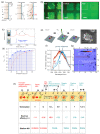
Energy band schematic of diamond (a). The 1.7 eV positive electron affinity with oxygen termination stabilizes the NV centers. On the contrary, hydrogen results in −1.0 eV electron affinity, transfer of electrons outside the surface, and the creation of a two-dimensional hole gas (2-DHG). The red line represents the NV instability while crossing the Fermi level close to the surface. (b) The FL contrast between hydrogen- and oxygen-terminated diamond (dark regions and bright squares, respectively) follows the concentration of NV centers. The contrast weakens with increasing nitrogen implantation dose, following a higher amount of donor impurities that reduce the negative impact of hydrogen at the surface. From Ref. [36], reprinted with permission. Surface oxidation (c) of nanodiamonds favors stabilization of NV, as evidenced by the reduction in the nondiamond sp2 signal and the lightening of the color of the solution. After this treatment, a NV fraction of up to 70% of the total can be obtained, even for small 10 nm particles (d). The red and blue colors correspond to the nonoxidized and oxidized NDs, respectively. Reprinted from ref. [93], with permission. A similar result was obtained via the selective fluorination of clustered NDs using electron irradiation in NF3 gas (e). Electron irradiation results in clusters of surface-fluorinated nanodiamonds. The PL of virgin, H-terminated (red curves), and fluorine-treated NDs (blue curves) is plotted in (f) together with the FL map separating the irradiated and the unirradiated regions (g). Reprinted from ref. [94], with permission. Types of terminations with surface electron affinity and stability of shallow NV are summarized in (h). Reprinted with permission from Kawai et al. [90]. Copyright 2019 ACS American Chemical Society.
Figure 5
Figure 5
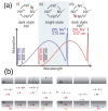
(a) Pictorial representation of the low-temperature absorption spectra of NV (red) and NV0 (blue). At the top, the relevant switching cycles are outlined. Arrows indicate laser-induced transition toward the excited states (denoted by the asterisk) or to a different charge state. A wavelength included in the gray-shaded region is able to excite and photoconvert both charge states; continuous charge switching is then ensured. In the white regions, the wavelength is not exciting either the NV (for λ < 450 nm) or the NV0 (for λ > 575 nm). In these cases, the photoconversion loop is broken (red crosses), and a preferential population of a particular charge state occurs. Reprinted from Ref. [40], with permission. (b) Schematic picture of charge conversion among neutral and negative NVs. NV to NV0 conversion involves two photons to detach an electron from the defect. The inverse process, NV0 to NV conversion, also occurs in two steps: an electron is excited to the e orbital from the a1 orbital in the band gap. The vacant place is then occupied by a second electron transferred from the deep-lying a1 orbital. The hole migrates away from the newly formed NV center. From Ref. [115], reprinted with permission.
Figure 6
Figure 6
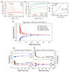
Several pulse sequences are employed to measure photoconversion. In (a), the ionization rate of shallow (<10 nm) NV is measured as a function of orange 594 nm laser power and duration (in ms). The colored dots denote the sequence of wavelengths of the pulsed lasers: a green laser initializes the NVs into the negative state, while the orange laser (of variable duration and power) reduces the NV fluorescence via photoionization to the neutral state. The red laser is used for read out. In (b), the recharge rate of NV is measured as a function of green 532 nm laser power and duration. The first two lasers (green and red) prepare the NV charge into the neutral state; the third laser (532 nm), of variable power and duration, restores the NV charge state, accompanying an increase in FL. The last pulse with a red laser is used to read out the charge state. Interestingly, ionization and recharge rates are quadratic with laser power for bulk NV but linear for shallow NV centers, suggesting a single-photon-mediated photoconversion mechanism (c). Adapted with permission from Dhomkar et al. [112]. Copyright 2018 American Chemical Society. The spin and charge dynamics can be decoupled using a magnetic field that mixes the spin statistics and quenches the FL (d). The signal arising from NV0 (blue lines) does not include any spin-related effect and is unaltered by the magnetic field. On the contrary, the quenching of FL is evident for the NV centers (red curves), suppressing the spin dynamics and revealing the charge dynamics. The NV0 and NV charge dynamics are complementary as they proceed in the opposite direction and can be isolated with a proper set of filters. The populations of NV and NV0 can be normalized and plotted together with their sum and ratio (e,f): it can be seen that after high-power laser irradiation, the NV/NV0 ratio remains large even after 100 ms. A possible explanation involves depleting the acceptor states by filling them with electrons released by substitutional nitrogen atoms, therefore stabilizing the negative NVs. Reprinted with permission from Gorrini et al. [35]. Copyright 2021 American Chemical Society.
Figure 7
Figure 7
Super-resolution imaging lead by charge photoconversion. In (a), the irradiation with red Gaussian-shaped (G) and green doughnut-shaped (D) laser beams emphasizes the NV emission, except in a small central region, where the NV centers are in the dark neutral state (the two photobleached spots at the center). In (b), a similar sequence with inverted laser wavelengths suppresses the FL everywhere (all the NV are neutral), except at the center (the two bright spots). By tuning the intensity and duration of the doughnut pulse, it is possible to improve the spatial resolution: this is evident when comparing traditional confocal imaging (c) with the present technique (d). For instance, points A and B, or C and D cannot be resolved individually with a confocal microscope, while super-resolution imaging allows extraction of fluorescence and ODMR signals. Readapted with permission from Chen et al. [18]. Copyright 2015 Springer Nature.
Figure 8
Figure 8
NV centers can serve as nanoprobes for the detection of magnetic and electric fields. A sophisticated setup constituted by an oxygen-terminated boron-doped single-crystal diamond (a) can initialize the NV center into the neutral state due to a combination of polyethylenimine (PEI) molecules electrostatically attached to the surface and an electrode to tune the electrostatic potential. When single-stranded DNA molecules adhere to the PEI layer, the surface electrostatic potential changes, and the NV centers turn to negative: it is possible to track this change in real time (b). In (c), the energy of the NV defects compared with Fermi energy (red line) is displayed for the two cases of PEI and PEI-DNA surface functionalization. In the pink-colored region, close to the oxygenated surface, the charge state of the NV centers depends on the presence of PEI or PEI-DNA molecules attached to the surface (dashed and solid black lines, respectively). Reprinted from Krečmarová et al. with permission [131]. Copyright 2021 American Chemical Society. When NDs are suspended with paramagnetic molecules (gadoteridol, in the figure), (d) the NV spin relaxes quicker and uncovers the signal due to charge dynamics, which proceeds in the opposite direction (e). The composition of spin relaxation and charge recovery produces a nonmonotonic trend that depends on the concentration of paramagnetic molecules per diamond nanoparticle. The net effect is a shift of the FL minimum, located at tmin, at shorter times (f). Readapted with permission from Gorrini et al. [26]. Copyright 2019 American Chemical Society.

Similar articles

References

    1. Kurtsiefer C., Mayer S., Zarda P., Weinfurter H. Stable solid-state source of single photons. Phys. Rev. Lett. 2000;85:290. doi: 10.1103/PhysRevLett.85.290. - DOI - PubMed
    1. Beveratos A., Kühn S., Brouri R., Gacoin T., Poizat J.P., Grangier P. Room temperature stable single-photon source. Eur. Phys. J. D-At. Mol. Opt. Plasma Phys. 2002;18:191–196. doi: 10.1140/epjd/e20020023. - DOI
    1. Babinec T.M., Hausmann B.J., Khan M., Zhang Y., Maze J.R., Hemmer P.R., Lončar M. A diamond nanowire single-photon source. Nat. Nanotechnol. 2010;5:195–199. doi: 10.1038/nnano.2010.6. - DOI - PubMed
    1. Schröder T., Gädeke F., Banholzer M.J., Benson O. Ultrabright and efficient single-photon generation based on nitrogen-vacancy centres in nanodiamonds on a solid immersion lens. New J. Phys. 2011;13:055017. doi: 10.1088/1367-2630/13/5/055017. - DOI
    1. Balasubramanian G., Neumann P., Twitchen D., Markham M., Kolesov R., Mizuochi N., Isoya J., Achard J., Beck J., Tissler J., et al. Ultralong spin coherence time in isotopically engineered diamond. Nat. Mater. 2009;8:383–387. doi: 10.1038/nmat2420. - DOI - PubMed