. 2023 Jun 30;13(7):698.
doi: 10.3390/bios13070698.

Multiplex PCR-Lateral Flow Dipstick Method for Detection of Thermostable Direct Hemolysin (TDH) Producing V. parahaemolyticus

Affiliations

Multiplex PCR-Lateral Flow Dipstick Method for Detection of Thermostable Direct Hemolysin (TDH) Producing V. parahaemolyticus

Jirakrit Saetang et al. Biosensors (Basel). .

Abstract

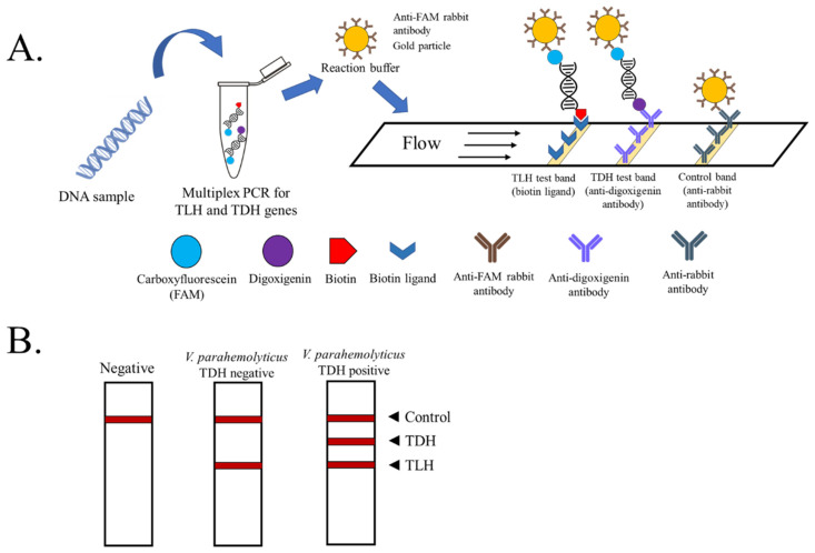
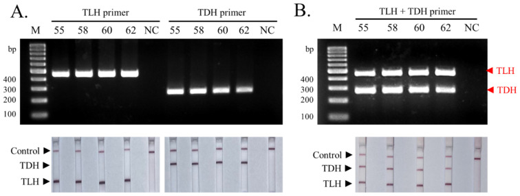
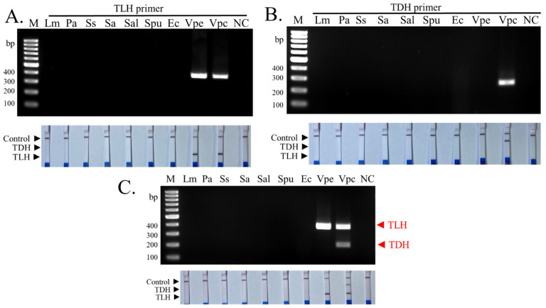
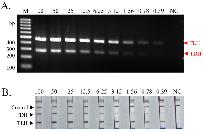
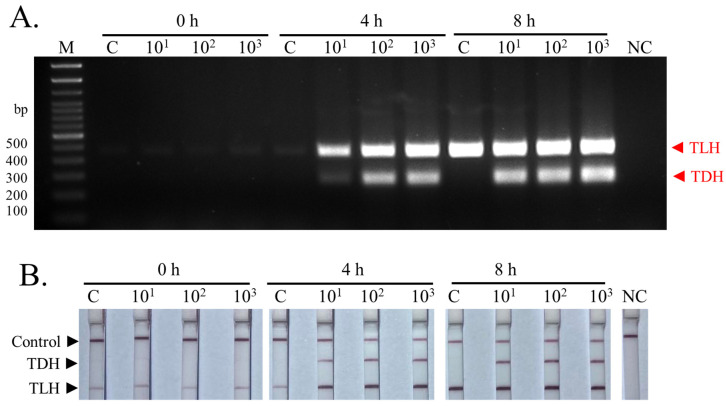
Vibrio parahaemolyticus is usually found in seafood and causes acute gastroenteritis in humans. Therefore, a detection method of pathogenic V. parahaemolyticus is necessary. Multiplex PCR combined with lateral flow dipstick (LFD) assay was developed to detect pathogenic V. parahaemolyticus. Biotin-, FAM-, and Dig-conjugated primers targeting thermolabile hemolysin (TLH) and thermostable direct hemolysin (TDH) genes were used for multiplex PCR amplification. The condition of the method was optimized and evaluated by agarose gel electrophoresis and universal lateral flow dipstick. The specificity assay was evaluated using strains belonging to seven foodborne pathogen species. The sensitivity of the method was also evaluated using DNA in the concentration range of 0.39-100 ng/reaction. The artificial spiking experiment was performed using 10 g of shrimp samples with an enrichment time of 0, 4, and 8 h with 101, 102, and 103 CFU of V. parahaemolyticus. The developed multiplex PCR-LFD assay showed no non-specific amplification with a limit of the detection of 0.78 ng DNA/reaction visualized by agarose gel electrophoresis and 0.39 ng DNA with LFD assay. The artificial spiking experiment demonstrated that this method could detect pathogenic V. parahaemolyticus at 10 CFU/10 g shrimp samples following a 4 h of enrichment. Multiplex PCR-LFD assay was therefore established for detecting pathogenic V. parahaemolyticus with high sensitivity and specificity and might be a useful tool to develop a detection kit used in the food safety sector.

Keywords: PCR; TDH; TLH; V. parahaemolyticus; lateral flow dipstick; seafood.


Conflict of interest statement

The authors declare no conflict of interest.

Figures

Figure 1
Figure 1
Schematic representation of the PCR product visualized by lateral flow assay (test strip from Milenia Biotec). (A) The principle of multiplex PCR-LFD for the detection and differentiation of pathogenic V. parahaemolyticus. (B) In the absence of amplicons of TLH and TDH, color appears at a control line only. If only TLH amplicon is found, the environmental strain of V. parahaemolyticus is presumed. The appearance of all three lines on the strip indicates the presence of pathogenic TDH+ V. parahaemolyticus in the sample.
Figure 2
Figure 2
Optimization of the annealing temperature for TLH and TDH primers using a DNA sample extracted from a pure culture of pathogenic V. parahaemolyticus. (A) The result of each annealing temperature on single primer PCR was visualized by agarose gel electrophoresis and LFD assay. (B) The result of each annealing temperature on multiplex PCR was visualized by agarose gel electrophoresis and LFD assay. The numbers 55, 58, 60, and 62 indicate the temperatures used for annealing. Lane M: 100 bp DNA marker. NC: negative control.
Figure 3
Figure 3
The specificity test of single primers. (A) The specificity of the TLH primer pairs against seven foodborne pathogenic bacteria visualized by agarose gel electrophoresis and LFD assay. (B) The specificity of the TDH primer pairs against seven foodborne pathogenic bacteria visualized by agarose gel electrophoresis and LFD assay. (C) The specificity of the multiplex PCR (TLH + TDH primers) against seven foodborne pathogenic bacteria was visualized by agarose gel electrophoresis and LFD assay. The experiments were done in duplicate. Lm: Listeria monocytogenes ATCC 15313, Pa: Pseudomonas aeruginosa ATCC 27853, Ss: Shigella sonnei PSU.SCB.16S.14, Sa: Staphylococcus aureus ATCC 25923, Sal: Shewanella sp. TBRC 5775, Spu: Shewanella putrefaciens JCM 20190, Ec: Escherichia coli ATCC 25922, Vpe: V. parahaemolyticus environmental strain, Vpc: V. parahaemolyticus clinical strain. NC: negative control.
Figure 4
Figure 4
Sensitivity test of the multiplex PCR-LFD assay for the identification of pathogenic V. parahaemolyticus. (A) The results visualized by agarose gel electrophoresis. (B) The results visualized by LFD assay. All concentrations of DNA used are nanograms (ng)/reaction. The experiments were done in duplicate. Lane M represents 100 bp DNA ladders. NC: negative control.
Figure 5
Figure 5
Limit of detection in spiked shrimp samples. The image shows the detection results of the multiplex PCR-LFD assay for spiked shrimp samples. The results were visualized by agarose gel electrophoresis (A) and LFD assay (B). The experiments were done in duplicate. Lane M represents 100 bp DNA ladders. NC: negative control.

Similar articles

Cited by

References

    1. Broberg C.A., Calder T.J., Orth K.V. parahaemolyticus Cell Biology and Pathogenicity Determinants. Microbes Infect. 2011;13:992–1001. doi: 10.1016/j.micinf.2011.06.013. - DOI - PMC - PubMed
    1. Yeung P.S.M., Boor K.J. Epidemiology, Pathogenesis, and Prevention of Foodborne V. parahaemolyticus Infections. Foodborne Pathog. Dis. 2004;1:74–88. doi: 10.1089/153531404323143594. - DOI - PubMed
    1. Ellett A.N., Rosales D., Jacobs J.M., Paranjpye R., Parveen S. Growth Rates of V. parahaemolyticus Sequence Type 36 Strains in Live Oysters and in Culture Medium. Microbiol. Spectr. 2022;10:e02112-22. doi: 10.1128/spectrum.02112-22. - DOI - PMC - PubMed
    1. Baker-Austin C., Trinanes J., Gonzalez-Escalona N., Martinez-Urtaza J. Non-Cholera Vibrios: The Microbial Barometer of Climate Change. Trends Microbiol. 2017;25:76–84. doi: 10.1016/j.tim.2016.09.008. - DOI - PubMed
    1. Wang R., Zhong Y., Gu X., Yuan J., Saeed A.F., Wang S. The Pathogenesis, Detection, and Prevention of V. parahaemolyticus. Front. Microbiol. 2015;6:144. doi: 10.3389/fmicb.2015.00144. - DOI - PMC - PubMed

LinkOut - more resources