Review
. 2023 Jul 2;8(3):284.
doi: 10.3390/biomimetics8030284.

Multifunctionality in Nature: Structure-Function Relationships in Biological Materials

Affiliations
Review

Multifunctionality in Nature: Structure-Function Relationships in Biological Materials

Jiaming Zhong et al. Biomimetics (Basel). .

Abstract

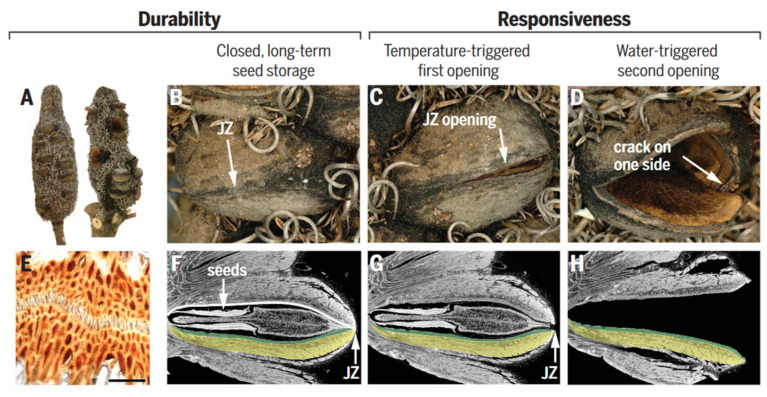
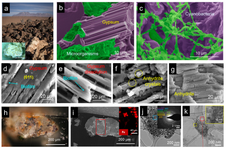
Modern material design aims to achieve multifunctionality through integrating structures in a diverse range, resulting in simple materials with embedded functions. Biological materials and organisms are typical examples of this concept, where complex functionalities are achieved through a limited material base. This review highlights the multiscale structural and functional integration of representative natural organisms and materials, as well as biomimetic examples. The impact, wear, and crush resistance properties exhibited by mantis shrimp and ironclad beetle during predation or resistance offer valuable inspiration for the development of structural materials in the aerospace field. Investigating cyanobacteria that thrive in extreme environments can contribute to developing living materials that can serve in places like Mars. The exploration of shape memory and the self-repairing properties of spider silk and mussels, as well as the investigation of sensing-actuating and sensing-camouflage mechanisms in Banksias, chameleons, and moths, holds significant potential for the optimization of soft robot designs. Furthermore, a deeper understanding of mussel and gecko adhesion mechanisms can have a profound impact on medical fields, including tissue engineering and drug delivery. In conclusion, the integration of structure and function is crucial for driving innovations and breakthroughs in modern engineering materials and their applications. The gaps between current biomimetic designs and natural organisms are also discussed.

Keywords: bioinspiration; biological materials; multifunctionality; multiscale structure; structure–function relationship.


Conflict of interest statement

The authors declare no conflict of interest.

Figures

Figure 1
Figure 1
Multifunction in natural organisms. Figures adapted from References [3,11,12,13,14,15,16,17,18,19].
Figure 2
Figure 2
Microstructures of functional tissues in representative natural organisms: the mantis shrimp dactyl club, the exoskeleton of a diabolical ironclad beetle, and chiton teeth. The impact of the mantis shrimp dactyl club last for a few milliseconds, while the crush of the beetle exoskeleton is close to a quasi-static state, and the teeth scratching is a long-time process subject to continuous loading. (A) HRTEM of HAP particles after heat treatment at 800 °C. (B) SEM micrograph of a transverse section of an intermolted dactyl club. Inset: nanoparticles ~60 nm in diameter are found within the impact surface. (C) Photograph of a mantis shrimp and its dactyl club, indicated with white arrows. (D) Snapshot of the hydrogen-bonding patterns in α-chitin after 10 ns of equilibration. Hydrogen bonds in blue are to nitrogen and red hydrogen bonds are between oxygen molecules. (E) False-colored SEM micrograph of fractured cross-section of the elytra, highlighting leaf-like setae (Se, green), epicuticle (Ep, red), exocuticle (Ex, yellow), endocuticle (En, blue), trabecula (Tr, orange), and hemolymph space (HS, violet). (F) Image of diabolical ironclad beetle scale bar. (G) HRTEM of the interface between a single-crystal domain and the mesocrystalline core; inset highlights a higher resolution of the boundary between the two domains. It shows a coherent interface between the single-crystalline domains located on the periphery of the mesocrystalline particles. (H) Piled rods observed at the tip of a fractured tooth, and an idealized hexagonal rod-like microstructure. (I) Radular teeth in chiton. (AC) adapted from Reference [13], (D) adapted from Reference [63], (E,F) adapted from Reference [14], (G) adapted from Reference [58], (H,I) adapted from Reference [64].
Figure 3
Figure 3
Structure–function relationships in spider silk. (A) Schematic of hierarchical and secondary structures. Amino acid motifs: A—Aniline; G—Glycine; P—Proline; X—random amino acid. (B) A shape memory structural model. (C) Net-point and switch model for moisture-sensitive shape memory mechanism. (A,B) adapted from Reference [72], (C) adapted from Reference [76].
Figure 4
Figure 4
Structure–function relationships in the mussel byssus. (A) Mussels attach to surfaces with a byssus. (B) The component structure of mussel byssal thread. (C) Schematic of the multiscale structure in the core of mussel byssal thread. (D) Idealized molecular model of byssal thread deformation and healing. (A,B) adapted from Reference [45], (C,D) adapted from Reference [12].
Figure 5
Figure 5
The formation process of plaque and structure–function relationships in the mussel. (A) The formation process of cavitation, pH adjustment, redox adjustment, and protein secretion. (B) Redox activity is driven by the difference between the high pH and O2 concentration of seawater versus the low pH and abundance of electron donors in the plaque. (C) Radial distribution of threads. (D) The spatulate geometry of a byssal thread and plaque. (E) The trabecular (spongy) structure of plaque. Figures adapted from Reference [11].
Figure 6
Figure 6
Structure and function in geckos. (A) Photo of a tokay gecko. (B) Photo of gecko toe pads. (C) Pictures showing multiscale structural hierarchy in gecko foot-hair. (D) Schematic illustration of structural compliance and adaptation against various rough surfaces. (E) Anti-fouling surfaces of gecko toe pads. (AC) adapted from Reference [95], (D,E) adapted from Reference [16].
Figure 7
Figure 7
Structure and function in Bogong moths. (A) A pinned specimen of a Bogong moth with exposed fore and hind wings. (B) Scanning electron micrographs of a forewing scale. Scale bars from left to right: 50 μm, 25 μm and 2.5 μm. (C) Bogong moth camouflage on the bark of the Argyle apple. Figures adapted from Reference [17].
Figure 8
Figure 8
Structure and function in chameleons. (A) Hematoxylin and eosin staining of a cross-section of white skin showing the epidermis (ep) and the two thick layers of iridophores. (B) TEM images of guanine nanocrystals in S-iridophores in the excited state and three-dimensional model of an FCC lattice. (C) TEM image of guanine nanocrystals in D-iridophores. (D) The overall color and (F) epidermal structure of Male m2 in a relaxed state (E). The overall color and (G) epidermal structure of Male m2 in an excited state. Scale bars, 20 mm (A); 200 nm (B,C,F,G). Figures adapted from Reference [18].
Figure 9
Figure 9
Structure and function of Banksia attenuata. (A,D) Cones from the North [(A), left side] contain mainly closed follicles (B). Half-open (C) and open follicles (D) are frequently found on cones in the South, where opening temperatures are lower [right infructescence in (A)]. (E) Light micrograph of a junction zone (JZ) sealed with wax (scale bar 100 mm). (F,H) Virtual cuts through micro-tomographic reconstructions of closed (F), half-open (G), and open follicles (H) showing the seeds with the separator in between [(F,G)] and the endocarp–mesocarp bilayer (colored in green and yellow in one of the two pericarp valves). The white line in (F) indicates the internal valve curvature, which changes with geographic location and climate. Figures adapted from Reference [3].
Figure 10
Figure 10
Cyanobacteria living in the Atacama Desert, Chile. (a) Gypsum rocks in the Atacama Desert. (b,c) SEM micrographs show cyanobacteria colonies in the gypsum rocks. (dg) The process of gypsum dissolution and phase transformation under the biofilm of the cyanobacteria colony. (h,i) Magnetite and hematite minerals were found in the gypsum rocks. (hj) TEM images show magnetite dissolution and phase transformation. (ag) adapted from Reference [19], (hk) adapted from Reference [112].

References

    1. Wegst U.G.K., Bai H., Saiz E., Tomsia A.P., Ritchie R.O. Bioinspired Structural Materials. Nat. Mater. 2015;14:23–36. doi: 10.1038/nmat4089. - DOI - PubMed
    1. Wang Y., Naleway S.E., Wang B. Biological and Bioinspired Materials: Structure Leading to Functional and Mechanical Performance. Bioact. Mater. 2020;5:745–757. doi: 10.1016/j.bioactmat.2020.06.003. - DOI - PMC - PubMed
    1. Eder M., Amini S., Fratzl P. Biological Composites-Complex Structures for Functional Diversity. Science. 2018;362:543–547. doi: 10.1126/science.aat8297. - DOI - PubMed
    1. Meyers M.A., Chen P.-Y., Lin A.Y.-M., Seki Y. Biological Materials: Structure and Mechanical Properties. Prog. Mater. Sci. 2008;53:1–206. doi: 10.1016/j.pmatsci.2007.05.002. - DOI - PubMed
    1. Meyers M.A., McKittrick J., Chen P.-Y. Structural Biological Materials: Critical Mechanics-Materials Connections. Science. 2013;339:773–779. doi: 10.1126/science.1220854. - DOI - PubMed

LinkOut - more resources