. 2023 Jul 23;9(7):592.
doi: 10.3390/gels9070592.

Development and Comparative Evaluation of Ciprofloxacin Nanoemulsion-Loaded Bigels Prepared Using Different Ratios of Oleogel to Hydrogels

Affiliations

Development and Comparative Evaluation of Ciprofloxacin Nanoemulsion-Loaded Bigels Prepared Using Different Ratios of Oleogel to Hydrogels

Rania Hamed et al. Gels. .

Abstract

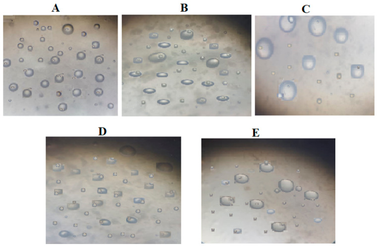
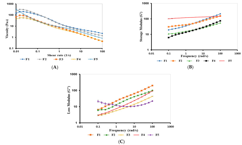
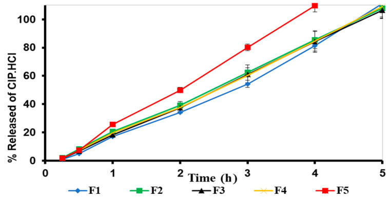
Nanoemulsions and bigels are biphasic delivery systems that can be used for topical applications. The aim of this study was to incorporate an oil-in-water ciprofloxacin hydrochloride nanoemulsion (CIP.HCl NE) into two types of bigels, Type I (oleogel (OL)-in-hydrogel (WH)) and Type II (WH-in-OL) to enhance drug penetration into skin and treat topical bacterial infections. Bigels were prepared at various ratios of OL and WH (1:1, 1:2, and 1:4). Initially, CIP.HCl NE was prepared and characterized in terms of droplet size, zeta potential, polydispersity index, morphology, and thermodynamic and chemical stability. Then CIP.HCl NE was dispersed into the OL or WH phase of the bigel. The primary physical stability studies showed that Type I bigels were physically stable, showing no phase separation. Whereas Type II bigels were physically unstable, hence excluded from the study. Type I bigels were subjected to microstructural, rheological, in vitro release, antimicrobial, and stability studies. The microscopic images showed a highly structured bigel network with nanoemulsion droplets dispersed within the bigel network. Additionally, bigels exhibited pseudoplastic flow and viscoelastic properties. A complete drug release was achieved after 4-5 h. The in vitro and ex vivo antimicrobial studies revealed that bigels exhibited antimicrobial activity against different bacterial strains. Moreover, stability studies showed that the rheological properties and physical and chemical stability varied based on the bigel composition over three months. Therefore, the physicochemical and rheological properties, drug release rate, and antimicrobial activity of Type I bigels could be modified by altering the OL to WH ratio and the phase in which the nanoemulsion dispersed in.

Keywords: antimicrobial activity; ciprofloxacin; hydrogel-in-oleogel bigel; nanoemulsions; oleogel-in-hydrogel bigel; rheology; topical delivery.


Conflict of interest statement

The authors declare no conflict of interest.

Figures

Figure 1
Figure 1
(A) Solubility of CIP.HCl in oils (OA and IPM), surfactants (TW-20 and Labrasol®), cosurfactants (PG, PEG, and ethanol), water, and 5% acetic acid. Data are presented as mean ± SD (n = 3), (B) CIP.HCl presents in different ionic forms based on the pH values (pH < 7, pH = 7, and pH > 7), (C) The interaction of CIP.HCl with 5% acetic acid, forming an acetate ion salt, and (D) a TEM image of CIP.HCl NE of spherical-shaped droplets.
Figure 1
Figure 1
(A) Solubility of CIP.HCl in oils (OA and IPM), surfactants (TW-20 and Labrasol®), cosurfactants (PG, PEG, and ethanol), water, and 5% acetic acid. Data are presented as mean ± SD (n = 3), (B) CIP.HCl presents in different ionic forms based on the pH values (pH < 7, pH = 7, and pH > 7), (C) The interaction of CIP.HCl with 5% acetic acid, forming an acetate ion salt, and (D) a TEM image of CIP.HCl NE of spherical-shaped droplets.
Figure 2
Figure 2
(A) Pictures of Type I bigels (F1–F5) showing no phase separation; (B) the range of yellow color of Type I bigels (F1–F5) which varied based on the proportion of OL; and (C) Type II bigels (F6–F10) showing phase separation and color change.
Figure 2
Figure 2
(A) Pictures of Type I bigels (F1–F5) showing no phase separation; (B) the range of yellow color of Type I bigels (F1–F5) which varied based on the proportion of OL; and (C) Type II bigels (F6–F10) showing phase separation and color change.
Figure 3
Figure 3
TEM images of bigels: (A) F1, (B) F2, (C) F3, (D) F4, and (E) F5.
Figure 4
Figure 4
Light-field micrographs of: (A) F1, (B) F2, (C) F3, (D) F4, and (E) F5 observed under 40× magnification.
Figure 5
Figure 5
FTIR spectra of (A) CIP.HCl, CP, and ET, and (B) PM, CIP.HCl NE, and F1–F5 bigels.
Figure 6
Figure 6
(A) Viscosity curves, (B) elastic G′ modulus of F1, F2, F3, F4, and F5 bigels, and (B) elastic G′ and (C) viscous G″ moduli of F1–F5 bigels. The data are presented as mean ± SD (n = 3).
Figure 7
Figure 7
In vitro release profiles of F1–F5 bigels through the cellulose membrane. The data are presented as the mean ± SD (n = 3).
Figure 8
Figure 8
Viscosity curves of (A) F1, (B) F2, (C) F3, (D) F4, and (E) F5 bigels after three months of storage at room temperature. The data are presented as mean ± SD (n = 3).
Figure 8
Figure 8
Viscosity curves of (A) F1, (B) F2, (C) F3, (D) F4, and (E) F5 bigels after three months of storage at room temperature. The data are presented as mean ± SD (n = 3).
Figure 9
Figure 9
Frequency-dependence (G′ and G″) of (A) F1, (B) F2, (C) F3, (D) F4, and (E) F5 bigels after three months of storage at room temperature. The data are presented as mean ± SD (n = 3).

Similar articles

Cited by

References

    1. Kahraman E., Kaykın M., Şahin Bektay H., Güngör S. Recent advances on topical application of ceramides to restore barrier function of skin. Cosmetics. 2019;6:52. doi: 10.3390/cosmetics6030052. - DOI
    1. Alkilani A.Z., Nasereddin J., Hamed R., Nimrawi S., Hussein G., Abo-Zour H., Donnelly R.F. Beneath the Skin: A Review of Current Trends and Future Prospects of Transdermal Drug Delivery Systems. Pharmaceutics. 2022;14:1152. doi: 10.3390/pharmaceutics14061152. - DOI - PMC - PubMed
    1. Tambunlertchai S., Geary S.M., Salem A.K. Skin penetration enhancement strategies used in the development of melanoma topical treatments. AAPS J. 2021;23:19. doi: 10.1208/s12248-020-00544-y. - DOI - PubMed
    1. Rehman K., Zulfakar M.H. Recent advances in gel technologies for topical and transdermal drug delivery. Drug Dev. Ind. Pharm. 2014;40:433–440. doi: 10.3109/03639045.2013.828219. - DOI - PubMed
    1. Souto E.B., Fangueiro J.F., Fernandes A.R., Cano A., Sanchez-Lopez E., Garcia M.L., Severino P., Paganelli M.O., Chaud M.V., Silva A.M. Physicochemical and biopharmaceutical aspects influencing skin permeation and role of SLN and NLC for skin drug delivery. Heliyon. 2022;8:e08938. doi: 10.1016/j.heliyon.2022.e08938. - DOI - PMC - PubMed

LinkOut - more resources