. 2023 Aug 31;14(4):e0082023.
doi: 10.1128/mbio.00820-23. Epub 2023 Jul 28.

SARS-CoV-2 infection alters mitochondrial and cytoskeletal function in human respiratory epithelial cells mediated by expression of spike protein

Affiliations

SARS-CoV-2 infection alters mitochondrial and cytoskeletal function in human respiratory epithelial cells mediated by expression of spike protein

Bonnie H Yeung-Luk et al. mBio. .

Abstract

Coronavirus disease 2019 (COVID-19) is caused by severe acute respiratory syndrome coronavirus 2 (SARS-CoV-2, SCV2), which has resulted in higher morbidity and mortality rate than other respiratory viral infections, such as Influenza A virus (IAV) infection. Investigating the molecular mechanisms of SCV2-host infection vs IAV is vital in exploring antiviral drug targets against SCV2. We assessed differential gene expression in human nasal cells upon SCV2 or IAV infection using RNA sequencing. Compared to IAV, we observed alterations in both metabolic and cytoskeletal pathways suggestive of epithelial remodeling in the SCV2-infected cells, reminiscent of pathways activated as a response to chronic injury. We found that spike protein interaction with the epithelium was sufficient to instigate these epithelial responses using a SCV2 spike pseudovirus. Specifically, we found downregulation of the mitochondrial markers SIRT3 and TOMM22. Moreover, SCV2 spike infection increased extracellular acidification and decreased oxygen consumption rate in the epithelium. In addition, we observed cytoskeletal rearrangements with a reduction in the actin-severing protein cofilin-1 and an increase in polymerized actin, indicating epithelial cytoskeletal rearrangements. This study revealed distinct epithelial responses to SCV2 infection, with early mitochondrial dysfunction in the host cells and evidence of cytoskeletal remodeling that could contribute to the worsened outcome in COVID-19 patients compared to IAV patients. These changes in cell structure and energetics could contribute to cellular resilience early during infection, allowing for prolonged cell survival and potentially paving the way for more chronic symptoms. IMPORTANCE COVID-19 has caused a global pandemic affecting millions of people worldwide, resulting in a higher mortality rate and concerns of more persistent symptoms compared to influenza A. To study this, we compare lung epithelial responses to both viruses. Interestingly, we found that in response to SARS-CoV-2 infection, the cellular energetics changed and there were cell structural rearrangements. These changes in cell structure could lead to prolonged epithelial cell survival, even in the face of not working well, potentially contributing to the development of chronic symptoms. In summary, these findings represent strategies utilized by the cell to survive the infection but result in a fundamental shift in the epithelial phenotype, with potential long-term consequences, which could set the stage for the development of chronic lung disease or long COVID-19.

Keywords: COVID-19; SARS-CoV-2; actin; bioenergetics; cytoskeleton; lung epithelia.


Conflict of interest statement

The authors declare no conflict of interest.

Figures

Fig 1
Fig 1
Transcriptomic profiling of human nasal cells with influenza A and SARS-CoV-2 infection. A (IAV) and SARS-CoV-2 (SCV2) infection. Venn diagrams (A and C) and heatmap (B and D) of differentially expressed genes in IAV and SCV2 vs mock at 12 hpi (A and B) and 24 hpi (C and D). Hierarchical clustering was performed to generate the heat maps. The color scale in the heatmaps corresponds to z-scores (standardized expression values). (E) The KEGG pathway enrichment analysis of the downregulated genes in SCV2 vs IAV at 24 hpi.
Fig 2
Fig 2
SCV2 decreases cell death signaling and promotes gene regulation. Using KEGG pathway analysis, differential expression of genes involved in (A) apoptosis, (B) necroptosis, (C) ferroptosis, and (D) gene regulation was shown in IAV/mock and SCV2/mock at 24 hpi.
Fig 3
Fig 3
SCV2 pseudovirus induces SCV2-specified gene expression in NHBE. NHBE cells were infected with SCV2 spike or control pseudovirus for 72 h, and (A) the infection efficiency was imaged by DsRed fluorescent signal (32 ×, scale bars of 100 µm). Gene expression of (B) HES1 and (C) KLF2 was examined by qPCR and normalized by housekeeping gene GAPDH. The error bars represent ±standard error of the mean (SEM) (n = 3). Statistics were determined by Welch’s test, with P < 0.05 considered statistically significant. GAPDH, glyceraldehyde-3-phosphate dehydrogenase; HES1, hes family BHLH transcription factor 1; KLF2, KLF transcription factor 2; NHBE, normal human bronchial epithelial; Spike, SARS-CoV-2 spike glycoprotein; VSV-G, VSV glycoprotein.
Fig 4
Fig 4
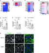
SCV2 infection enhances metabolism in the host cells. Using KEGG pathway analysis, differentially expressed genes involved in (A) nucleotide, (B) amino acid, (C) fatty acid, and carbohydrate metabolisms were shown in IAV/mock and SCV2/mock at 24 hpi. After 72 h of pseudovirus infection in NHBE cells, gene expression of (E) glycolysis rate limiting PKLR, (F) SIRT3, and (G) mitochondrial marker TOMM22 was measured by qPCR and normalized by housekeeping gene GAPDH (5–9 inserts from two donors), and (H) the mitochondrial localization was observed by TOMM22 immunofluorescent staining. The immunofluorescence images were taken at 63× oil objective, scale bars of 20 µm. The error bars represent ±standard error of the mean (SEM) (two donors). Statistics were determined by Welch’s test, with P < 0.05 considered statistically significant. GAPDH: glyceraldehyde-3-phosphate dehydrogenase. PKLR, pyruvate kinase L/R; SIRT3, NAD+-dependent protein deacetylases 3; TOMM22, translocase of the outer membrane of mitochondria; Spike, SARS-CoV-2 spike glycoprotein; VSV-G, VSV glycoprotein.
Fig 5
Fig 5
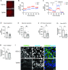
SCV2 spike protein infection impairs metabolism in human epithelial cells. (A) The pseudovirus infection efficiency of Calu3 cells after 48 h post infection was examined by DsRed fluorescent signal (32×, scale bars of 100 µm). (B) ECAR and (C) OCR were examined in the pseudovirus-infected Calu3 cells. (D) Basal respiration, (E) maximal respiration, and (F) ATP-linked respiration were shown. Gene expression of (G) SIRT3 and (H) TOMM22 as well as (I) immunofluorescence of TOMM22 (green) and Hoechst (blue) staining were taken at 63× oil objective, scale bar of 20 µm. The error bars represent ±standard error of the mean (SEM) (4–10 inserts/group). Statistics were determined by Welch’s test, with P < 0.05 considered statistically significant. ECAR, extracellular acidification rate; OCR, oxygen consumption rate; RHOB, ras homolog family member B; SIRT3, NAD+-dependent protein deacetylases 3; TOMM22, translocase of the outer membrane of mitochondria; Spike, SARS-CoV-2 spike glycoprotein; VSV-G, VSV glycoprotein.
Fig 6
Fig 6
SCV2 infection disrupts cell-cell adhesion in the human epithelium. Using KEGG pathway analysis, differential expression of genes involved in (A) tight junction, (B) cell adhesion, (C) focal adhesion, and (D) regulation of actin cytoskeleton was shown in IAV/mock and SCV2/mock at 24 hpi. After 72 h of the pseudovirus infection in NHBE cells, expressions of (E) CFL1 transcript, (F) CFL1 protein, and (G) RHOB transcript were examined (n = 3–12). (G) Immunofluorescence of TOMM22 (green) and Hoechst (blue) staining was taken at 63× oil objective, scale bar of 10 µm. TEER was measured in the pseudovirus-infected (H) NHBE cells and (I) Calu3 cells (n = 8). The error bars represent ±standard error of the mean (SEM). Statistics were determined by Welch’s test, with P < 0.05 considered statistically significant. CFL1, cofilin-1; ECAR, extracellular acidification rate; GAPDH, glyceraldehyde-3-phosphate dehydrogenase; OCR, oxygen consumption rate; TEER, transepithelial electrical resistance; TOMM22, translocase of the outer membrane of mitochondria; Spike, SARS-CoV-2 spike glycoprotein; VSV-G, VSV glycoprotein.
Fig 7
Fig 7
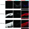
F-actin is increased in human lungs with COVID-19. Immunofluorescent staining of phalloidin (red) and Hoechst (blue) was performed in human lungs from normal and COVID-19 patients. The immunofluorescence images were taken at 63× oil objective (scale bars of 10 µm).

References

    1. Guan W-J, Ni Z-Y, Hu Y, Liang W-H, Ou C-Q, He J-X, Liu L, Shan H, Lei C-L, Hui DSC, Du B, Li L-J, Zeng G, Yuen K-Y, Chen R-C, Tang C-L, Wang T, Chen P-Y, Xiang J, Li S-Y, Wang J-L, Liang Z-J, Peng Y-X, Wei L, Liu Y, Hu Y-H, Peng P, Wang J-M, Liu J-Y, Chen Z, Li G, Zheng Z-J, Qiu S-Q, Luo J, Ye C-J, Zhu S-Y, Zhong N-S. 2020. Clinical characteristics of coronavirus disease 2019 in China. N Engl J Med 382:1708–1720. doi:10.1056/NEJMoa2002032 - DOI - PMC - PubMed
    1. Huang C, Wang Y, Li X, Ren L, Zhao J, Hu Y, Zhang L, Fan G, Xu J, Gu X, Cheng Z, Yu T, Xia J, Wei Y, Wu W, Xie X, Yin W, Li H, Liu M, Xiao Y, Gao H, Guo L, Xie J, Wang G, Jiang R, Gao Z, Jin Q, Wang J, Cao B. 2020. Clinical features of patients infected with 2019 novel coronavirus in Wuhan, China. Lancet 395:497–506. doi:10.1016/S0140-6736(20)30183-5 - DOI - PMC - PubMed
    1. Hoffmann M, Kleine-Weber H, Schroeder S, Krüger N, Herrler T, Erichsen S, Schiergens TS, Herrler G, Wu NH, Nitsche A, Müller MA, Drosten C, Pöhlmann S. 2020. SARS-CoV-2 cell entry depends on ACE2 and TMPRSS2 and is blocked by a clinically proven protease inhibitor. Cell 181:271–280. doi:10.1016/j.cell.2020.02.052 - DOI - PMC - PubMed
    1. Letko M, Marzi A, Munster V. 2020. Functional assessment of cell entry and receptor usage for SARS-CoV-2 and other lineage B betacoronaviruses. Nat Microbiol 5:562–569. doi:10.1038/s41564-020-0688-y - DOI - PMC - PubMed
    1. Dou D, Revol R, Östbye H, Wang H, Daniels R. 2018. Influenza A virus cell entry, replication, virion assembly and movement. Front Immunol 9:1581. doi:10.3389/fimmu.2018.01581 - DOI - PMC - PubMed

Publication types