Review
. 2023 Jun 30;14(7):349.
doi: 10.3390/jfb14070349.

Performance-Enhancing Materials in Medical Gloves

Affiliations
Review

Performance-Enhancing Materials in Medical Gloves

María José Lovato et al. J Funct Biomater. .

Abstract

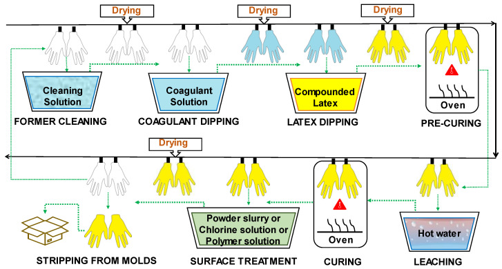
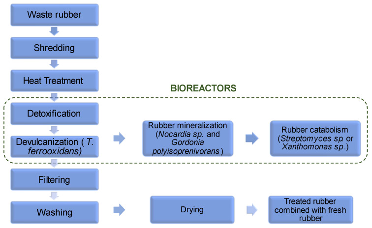
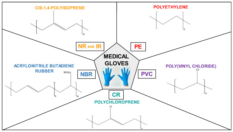
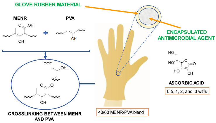
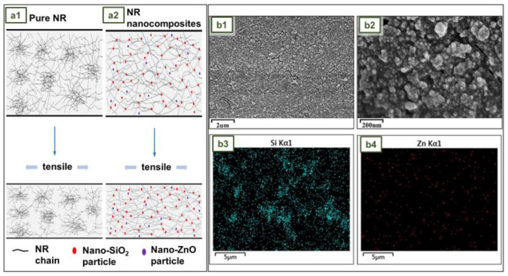
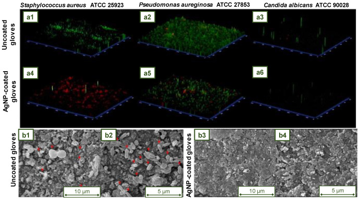
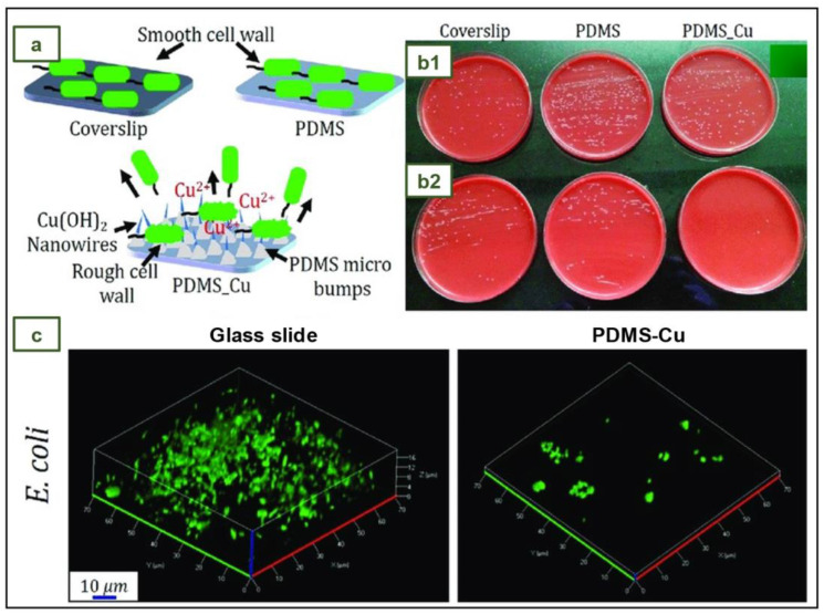
Medical gloves, along with masks and gowns, serve as the initial line of defense against potentially infectious microorganisms and hazardous substances in the health sector. During the COVID-19 pandemic, medical gloves played a significant role, as they were widely utilized throughout society in daily activities as a preventive measure. These products demonstrated their value as important personal protection equipment (PPE) and reaffirmed their relevance as infection prevention tools. This review describes the evolution of medical gloves since the discovery of vulcanization by Charles Goodyear in 1839, which fostered the development of this industry. Regarding the current market, a comparison of the main properties, benefits, and drawbacks of the most widespread types of sanitary gloves is presented. The most common gloves are produced from natural rubber (NR), polyisoprene (IR), acrylonitrile butadiene rubber (NBR), polychloroprene (CR), polyethylene (PE), and poly(vinyl chloride) (PVC). Furthermore, the environmental impacts of the conventional natural rubber glove manufacturing process and mitigation strategies, such as bioremediation and rubber recycling, are addressed. In order to create new medical gloves with improved properties, several biopolymers (e.g., poly(vinyl alcohol) and starch) and additives such as biodegradable fillers (e.g., cellulose and chitin), reinforcing fillers (e.g., silica and cellulose nanocrystals), and antimicrobial agents (e.g., biguanides and quaternary ammonium salts) have been evaluated. This paper covers these performance-enhancing materials and describes different innovative prototypes of gloves and coatings designed with them.

Keywords: antimicrobial properties; bio-filler; medical gloves; natural rubber; performance-enhancing materials; reinforcing filler; synthetic rubber.


Conflict of interest statement

The authors declare no conflict of interest.

Figures

Figure 1
Figure 1
Imports of surgical gloves in the EU-27 from January 2019 to December 2021. Chart prepared by the authors based on Eurostat data [16].
Figure 2
Figure 2
Exports of rubber gloves from Malaysia. The arrow indicates the sharp rise. Chart prepared by the authors based on MARGMA data shown in reference [18].
Figure 3
Figure 3
Quarterly financial report of Top Glove Corporation Berhad. Chart prepared by the authors based on Bursa Malaysia data shown in reference [21].
Figure 4
Figure 4
Quarterly financial report of Comfort Gloves Berhad. Chart prepared by the authors based on Bursa Malaysia data shown in reference [22].
Figure 5
Figure 5
Production process of medical gloves by dipping.
Figure 6
Figure 6
Recycling and remediation of NR through microbial action. Reprinted and adapted with permission from reference [46]. Copyright © 2013 Springer Nature.
Figure 7
Figure 7
Chemical structure of common types of medical gloves.
Figure 8
Figure 8
MENR/PVA blend glove with encapsulated AA. Graphic prepared by the authors based on reference information [83].
Figure 9
Figure 9
SEM images of (a1) NSS and (a2) AHSS. The red circle shows the porous surface of the starch particle after acid hydrolysis (b) Mass loss of NR and XNBR films (control, NSS-filled, and AHSS-filled) after 3 weeks. Reprinted and adapted with permission from reference [34]. Copyright © 2019 Elsevier.
Figure 10
Figure 10
Starch hydrolysis test of the mixture culture. The blue and the orange arrows show the areas where the starch remains unchanged and where it has been hydrolyzed to glucose by microbial action, respectively. Reprinted and adapted with permission under a Creative Commons license (CC BY 3.0) from reference [35].
Figure 11
Figure 11
SEM (a1,a2) and FESEM (b1b4) micrographs of mangosteen peel. The main physical properties of mangosteen peel powder are summarized below. Reprinted and adapted with permission from reference [93]. Copyright © 2020 John Wiley and Sons.
Figure 12
Figure 12
(a) Depolymerization of cellulose to nanocellulose (reprinted with permission under a Creative Commons license (CC BY 3.0) from reference [96]). (b) Illustration of the formation of a Zn–cellulose complex with CNC in the cross-linked NR matrix [99]. (c) Illustration of the proposed permeation mechanism through NR and NR–CNC nanocomposites and THF. (b,c) Reprinted and adapted with permission from reference [99]. Copyright © 2020 American Chemical Society.
Figure 13
Figure 13
(a1) Brief exposure test of Gardine-coated gloves. (a2) Long-term exposure. (b) Mean colony counts recorded for all coated glove types after 24 h exposure to MRSA or E. coli. Reprinted and adapted with permission from reference [101]. Copyright © 2009 Elsevier.
Figure 14
Figure 14
(a) Schematic illustration of coating (illustration prepared by the authors based on reference information [105]). (b) Pre- and post-exposure populations of challenge microorganisms following transfer procedures. Adapted with permission under a Creative Commons license (CC BY) from reference [105]. Copyright © 2013 Elsevier.
Figure 15
Figure 15
Three-layer NR glove with antimicrobial agent. (a) Cross-section micrograph. (b) Three-layer scheme. Reprinted with permission from reference [106]. Copyright © 2011 Elsevier.
Figure 16
Figure 16
(a) Mode of action of QACs against both bacterial and viral phospholipid membranes (Reprinted with permission under standard ACS Author Choice/Editors’ Choice usage agreement from reference [110]). (b) Antibacterial activity of QP-4VP-conjugated NR films vs. Control NR films. Reprinted with permission from reference [111]. Copyright © 2022 Elsevier.
Figure 17
Figure 17
(a1,a2) Schematic diagram of nanoparticle enhancement mechanism. Scanning electron microscopy images of (b1,b2) LZ1S4.2 and element distribution of (b3) Si and (b4) Zn. Reprinted with permission from reference [114]. Copyright © 2022 John Wiley and Sons.
Figure 18
Figure 18
Fluorescence microscopy images of (a1,a4) S. aureus ATCC 25923, (a2,a5) P. aeruginosa ATCC 27853, and (a3,a6) C. albicans ATCC 90028 biofilms incubated with (a1a3) uncoated gloves and (a4a6) AgNP-coated gloves. Scanning electron microscopy (SEM) analysis. SEM micrograph of polymicrobial anti biofilm activity of (b1,b2) uncoated gloves and (b3,b4) AgNP-coated gloves. Reprinted with permission from reference [121]. Copyright © 2021 John Wiley and Sons.
Figure 19
Figure 19
Images of the antibacterial activity of PDMS-Cu. Reprinted with permission from reference [126]. Copyright © 2018 American Chemical Society.

References

    1. Mazón L., Orriols R.M. Sanitary Gloves Management. Adequate Protection-Effectiveness and Environmental Responsibility. Rev. Asoc. Española Espec. Med. Trab. 2018;27:175–181.
    1. Delves P.J., Martin S.J., Burton D.R., Roitt I.M. Roitt’s Essential Immunology. 13th ed. John Wiley and Sons; Hoboken, NJ, USA: 2017.
    1. Ford C., Park L.J. How to Apply and Remove Gloves. Br. J. Nurs. 2019;28:26–28. doi: 10.12968/bjon.2019.28.1.26. - DOI - PubMed
    1. Babadi A.A., Bagheri S., Hamid S.B.A. Progress on Antimicrobial Surgical Gloves: A Review. Rubber Chem. Technol. 2016;89:117–125. doi: 10.5254/rct.15.84882. - DOI
    1. Ellis H. Surgical Gloves. J. Perioper. Pract. 2010;20:219–220. doi: 10.1177/175045891002000606. - DOI - PubMed

LinkOut - more resources