. 2023 Jul 3;21(7):391.
doi: 10.3390/md21070391.

Protective Effect of Fucoxanthin on Zearalenone-Induced Hepatic Damage through Nrf2 Mediated by PI3K/AKT Signaling

Affiliations

Protective Effect of Fucoxanthin on Zearalenone-Induced Hepatic Damage through Nrf2 Mediated by PI3K/AKT Signaling

Rebai Ben Ammar et al. Mar Drugs. .

Abstract

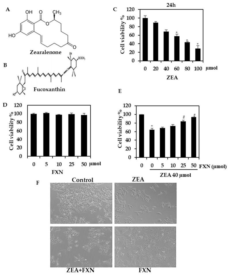
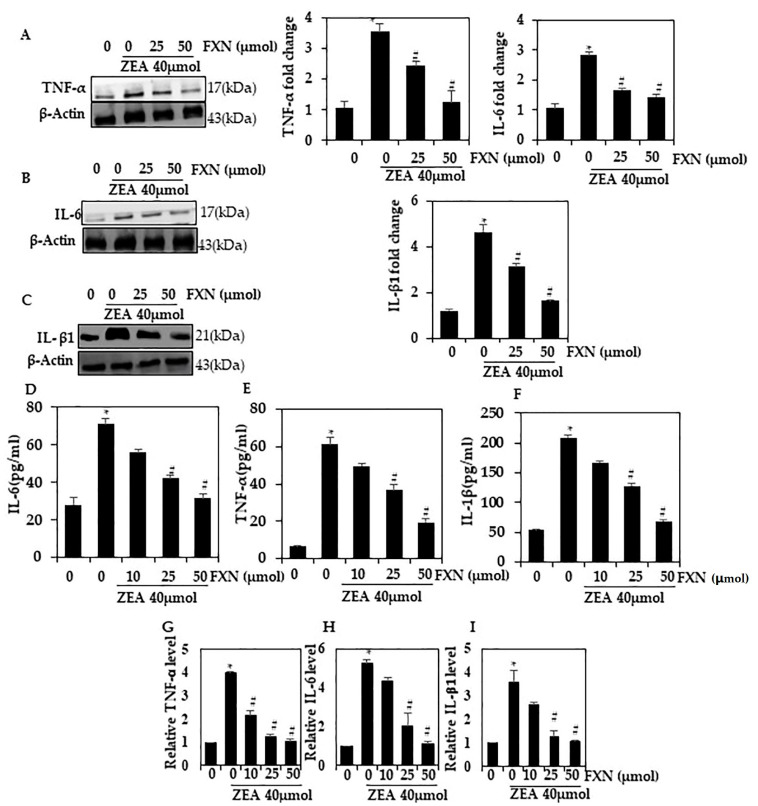
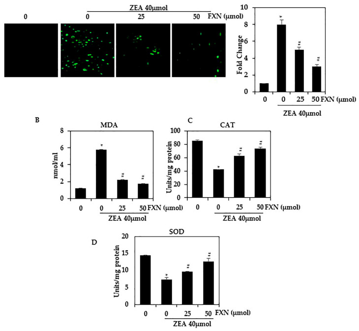
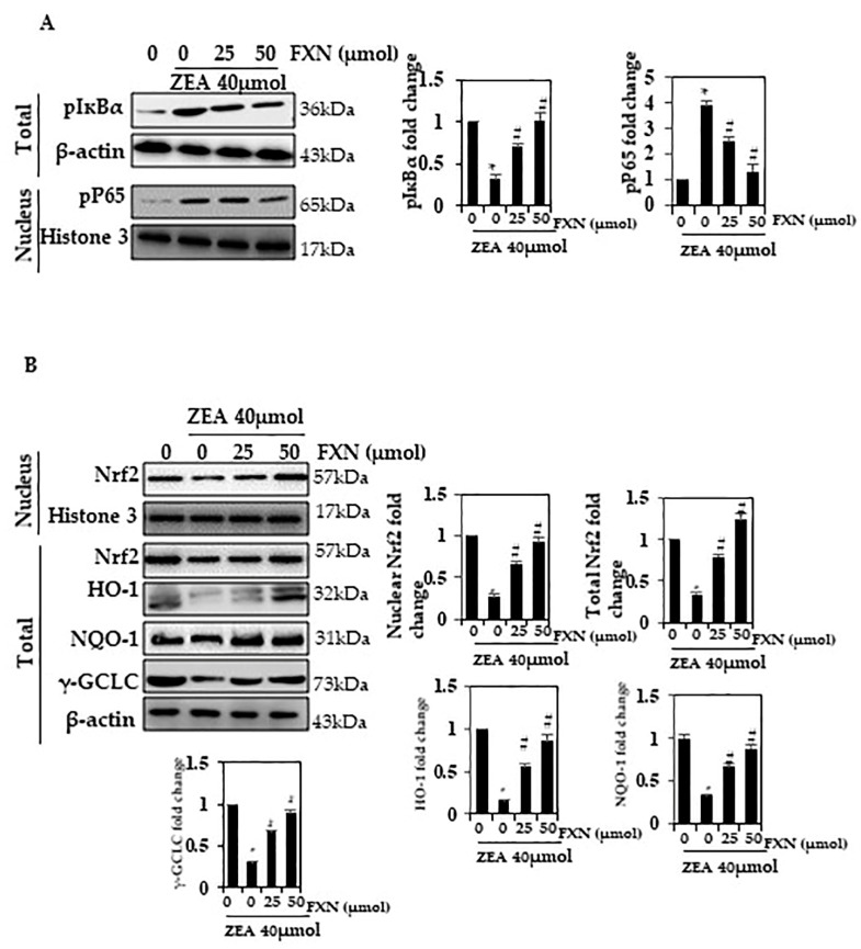
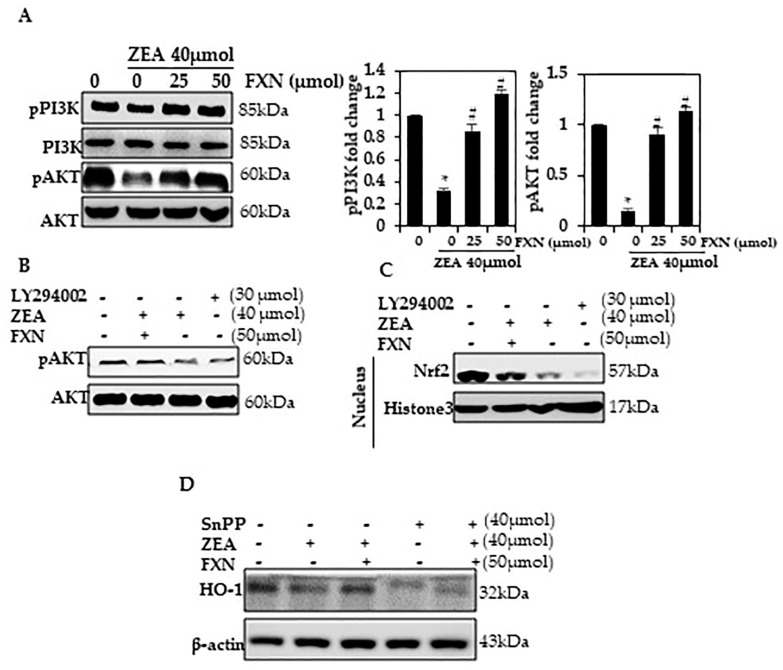
Hepatotoxic contaminants such as zearalenone (ZEA) are widely present in foods. Marine algae have a wide range of potential applications in pharmaceuticals, cosmetics, and food products. Research is ongoing to develop treatments and products based on the compounds found in algae. Fucoxanthin (FXN) is a brown-algae-derived dietary compound that is reported to prevent hepatotoxicity caused by ZEA. This compound has multiple biological functions, including anti-diabetic, anti-obesity, anti-microbial, and anti-cancer properties. Furthermore, FXN is a powerful antioxidant. In this study, we examined the effects of FXN on ZEA-induced stress and inflammation in HepG2 cells. MTT assays, ROS generation assays, Western blots, and apoptosis analysis were used to evaluate the effects of FXN on ZEA-induced HepG2 cell inflammation. Pre-incubation with FXN reduced the cytotoxicity of ZEA toward HepG2 cells. FXN inhibited the ZEA-induced production of pro-inflammatory cytokines, including IL-1 β, IL-6, and TNF-α. Moreover, FXN increased HO-1 expression in HepG2 by activating the PI3K/AKT/NRF2 signaling pathway. In conclusion, FXN inhibits ZEA-induced inflammation and oxidative stress in hepatocytes by targeting Nrf2 via activating PI3K/AKT signaling.

Keywords: HepG2; Nrf2; PI3K/AKT; fucoxanthin; marine algae; oxidative stress.


Conflict of interest statement

The authors declare no conflict of interest.

Figures

Figure 1
Figure 1
Analysis of ZEA and FXN viability. (A,B) Chemical structure of ZEA and FXN. (C) The viability of HepG2 cells was determined using the MTT assay after the addition of the indicated ZEA dosages for 24 h. (D) FXN cytotoxicity to HepG2 cells. (E) In the MTT assay, FXN protects against ZEA cytotoxicity (24 h). (F) HepG2 morphological changes. Values are presented as the mean and standard error of the mean (SD). One-way analysis of variance followed by Tukey’s post hoc test analysis was used for inter-group comparison. A significant difference is defined as a difference between the mean and standard deviation of three duplicates (n = 3), with * p < 0.05 control vs. ZEA and # p < 0.0.5ZEA vs. pre-treated.
Figure 2
Figure 2
Hepatic cells were protected from ZEA-induced cytokine production by FXN. FXN and/or ZEA were administered to HepG2 cells for 24 h. Western blotting was used to assess the expression of TNF-α, IL-6, and IL-β1 proteins in (AC). (DF) IL-6, IL-1β, TNF-α, and VCAMsecretion into the culture media was quantified using a commercial ELISA kit. (GI) mRNA level of the pro-inflammatory cytokines TNF-α, IL-6, and ILβ1 by RT-PCR. Values are presented as the mean and standard error of the mean (SD). One-way analysis of variance followed by Tukey’s post hoc test analysis was used for inter-group comparison. A significant difference is defined as a difference between the mean and standard deviation of three duplicates (n = 3), with * p < 0.05 control vs. ZEA and # p < 0.05 ZEA vs. pre-treated.
Figure 3
Figure 3
Effect of FXN on ZEA-induced ROS production in HepG2 cells. As shown in (A), HepG2 cells were then pre-treated with FXN (0, 25, and 50 µM) for 2 h, followed by ZEA (40 µM) for 4 h. H2DCFDA fluorescence was used to measure intracellular ROS levels. (BD) MDA, SOD, and CAT. Values are presented as the mean and standard error of the mean (SD). One-way analysis of variance followed by Tukey’s post hoc test analysis was used for inter-group comparison. A significant difference is defined as a difference between the mean and standard deviation of three duplicates (n = 3), with * p < 0.05 control vs. ZEA and # p < 0.05 ZEA vs. pre-treated.
Figure 4
Figure 4
An altered FXN response to NF-кB p65 activation. (A) Nuclear protein extract and total protein extract were detected using p-IкBα and pNF-кB p65 antibodies on SDS-PAGE. (B) Western blot analysis was conducted to determine nuclear Nrf2, and total NQO-1, HO-1, and γ-GCLC. Values are presented as the mean and standard error of the mean (SD). One-way analysis of variance followed by Tukey’s post hoc test analysis was used for inter-group comparison. A significant difference is defined as a difference between the mean and standard deviation of three duplicates (n = 3), with * p < 0.05 control vs. ZEA and # p < 0.05 ZEA vs. pre-treated.
Figure 5
Figure 5
PI3K/AKT phosphorylation is activated by FXN. (A) The cells were treated with ZEA or FXN for 24 h. Western blotting results list the relative ratios of PI3K and AKT expression. (B,C) The cells were pre-treated with PI3K/AKT inhibitors for 2 h and then treated with FXN with or without ZEA for 24 h. Western blotting detected pAKT and Nrf2. (D) A Western blot was conducted after cells were exposed to SnPP for 1 h, followed by treatment with ZEA and FXN for 24 h. Values are presented as the mean and standard error of the mean (SD). One-way analysis of variance followed by Tukey’s post hoc test analysis was used for inter-group comparison. A significant difference is defined as a difference between the mean and standard deviation of three duplicates (n = 3), with * p < 0.05 control vs. ZEA and # p < 0.05 ZEA vs. pre-treated.

References

    1. Marin D., Pistol G., Bulgaru C., Taranu I. Cytotoxic and inflammatory effects of individual and combined exposure of HepG2 cells to zearalenone and its metabolites. Naunyn-Schmiedeberg’s Arch. Pharmacol. 2019;392:937–947. doi: 10.1007/s00210-019-01644-z. - DOI - PubMed
    1. Han X., Huangfu B., Xu T., Xu W., Asakiya C., Huang K., He X. Research progress of safety of zearalenone: A review. Toxins. 2022;14:386. doi: 10.3390/toxins14060386. - DOI - PMC - PubMed
    1. Feng Y.-Q., Zhao A.-H., Wang J.-J., Tian Y., Yan Z.-H., Dri M., Shen W., De Felici M., Li L. Oxidative stress as a plausible mechanism for zearalenone to induce genome toxicity. Gene. 2022;829:146511. doi: 10.1016/j.gene.2022.146511. - DOI - PubMed
    1. Rajendran P., Ammar R.B., Al-Saeedi F.J., Mohamed M.E., ElNaggar M.A., Al-Ramadan S.Y., Bekhet G.M., Soliman A.M. Kaempferol inhibits zearalenone-induced oxidative stress and apoptosis via the PI3K/Akt-mediated Nrf2 signaling pathway: In vitro and in vivo studies. Int. J. Mol. Sci. 2021;22:217. doi: 10.3390/ijms22010217. - DOI - PMC - PubMed
    1. Wu D., Zhong P., Wang Y., Zhang Q., Li J., Liu Z., Ji A., Li Y. Hydrogen sulfide attenuates high-fat diet-induced non-alcoholic fatty liver disease by inhibiting apoptosis and promoting autophagy via reactive oxygen spe-cies/phosphatidylinositol 3-kinase/AKT/mammalian target of rapamycin signaling pathway. Front. Pharmacol. 2020;11:585860. doi: 10.3389/fphar.2020.585860. - DOI - PMC - PubMed

LinkOut - more resources