Review
. 2023 Dec;19(12):3033-3061.
doi: 10.1080/15548627.2023.2238577. Epub 2023 Jul 28.

The constructive and destructive impact of autophagy on both genders' reproducibility, a comprehensive review

Affiliations
Review

The constructive and destructive impact of autophagy on both genders' reproducibility, a comprehensive review

Mohammad Samare-Najaf et al. Autophagy. 2023 Dec.

Erratum in

  • Correction.
    [No authors listed] [No authors listed] Autophagy. 2025 Jun;21(6):iv-vi. doi: 10.1080/15548627.2023.2257071. Epub 2023 Sep 13. Autophagy. 2025. PMID: 37700720 Free PMC article. No abstract available.

Abstract

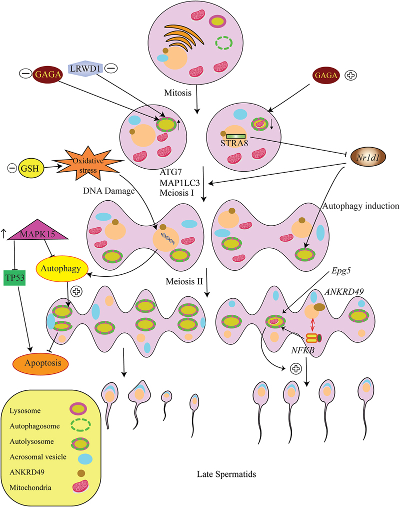
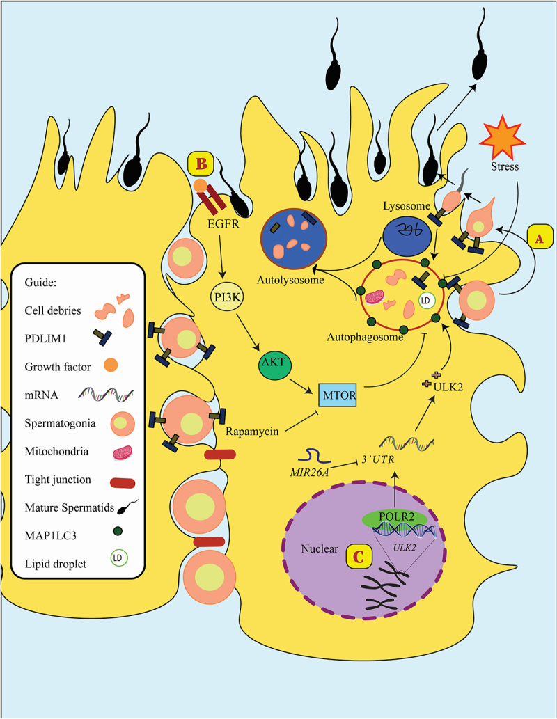
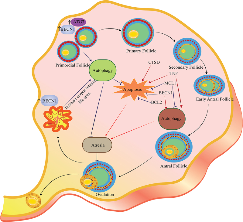
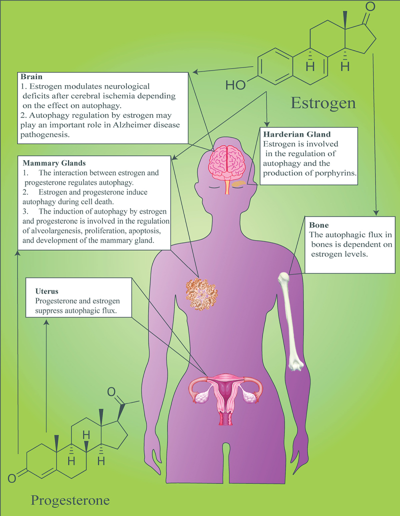
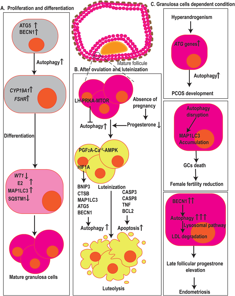
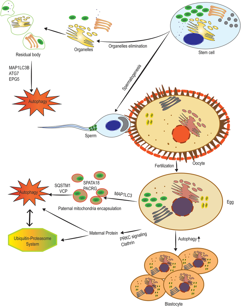
Reproduction is characterized by a series of massive renovations at molecular, cellular, and tissue levels. Recent studies have strongly tended to reveal the involvement of basic molecular pathways such as autophagy, a highly conserved eukaryotic cellular recycling, during reproductive processes. This review comprehensively describes the current knowledge, updated to September 2022, of autophagy contribution during reproductive processes in males including spermatogenesis, sperm motility and viability, and male sex hormones and females including germ cells and oocytes viability, ovulation, implantation, fertilization, and female sex hormones. Furthermore, the consequences of disruption in autophagic flux on the reproductive disorders including oligospermia, azoospermia, asthenozoospermia, teratozoospermia, globozoospermia, premature ovarian insufficiency, polycystic ovarian syndrome, endometriosis, and other disorders related to infertility are discussed as well.Abbreviations: AKT/protein kinase B: AKT serine/threonine kinase; AMPK: AMP-activated protein kinase; ATG: autophagy related; E2: estrogen; EDs: endocrine disruptors; ER: endoplasmic reticulum; FSH: follicle stimulating hormone; FOX: forkhead box; GCs: granulosa cells; HIF: hypoxia inducible factor; IVF: in vitro fertilization; IVM: in vitro maturation; LCs: Leydig cells; LDs: lipid droplets; LH: luteinizing hormone; LRWD1: leucine rich repeats and WD repeat domain containing 1; MAP1LC3: microtubule associated protein 1 light chain 3; MAPK: mitogen-activated protein kinase; MTOR: mechanistic target of rapamycin kinase; NFKB/NF-kB: nuclear factor kappa B; P4: progesterone; PCOS: polycystic ovarian syndrome; PDLIM1: PDZ and LIM domain 1; PI3K: phosphoinositide 3-kinase; PtdIns3P: phosphatidylinositol-3-phosphate; PtdIns3K: class III phosphatidylinositol 3-kinase; POI: premature ovarian insufficiency; ROS: reactive oxygen species; SCs: Sertoli cells; SQSTM1/p62: sequestosome 1; TSGA10: testis specific 10; TST: testosterone; VCP: vasolin containing protein.

Keywords: Apoptosis; autophagy; fertility; follicles; granulosa cells; infertility.


Conflict of interest statement

No potential conflict of interest was reported by the author(s).

Figures

Figure 1.
Figure 1.
The role of autophagy during spermatogenesis. The induced overproduction of GAGA in the second or third mitosis in early spermatogenesis leads to an increase in the presence of autophagosomes, an increased autophagic flux, and a significant increase in mitophagy in spermatogonia. Moreover, the expression of Epg5 is involved in the augmentation of autophagy and mitochondrial clearance during spermatogenesis. Interestingly, the Nr1d1-mediated suppression of autophagy by STRA8 is necessary for mitotic to miotic transition. The rate of autophagy in spermatogenesis increases after meiosis I. Conversely, decreased levels of GSH increase the levels of MAP1LC3 proteins showing that autophagy is induced under oxidative stress, leading to the development and survival of male germ cells with more DNA damage and sperm deformations followed by azoospermia and oligozoospermia. ANKRD49, along with the NFKB pathway, increases the spermatogenesis rate via autophagy-dependent survival. In addition, overexpression of MAPK15 disrupts autophagy supportive function that controls the prevention of DNA damage and the activation of the TP53, hence causing malignant transformation of germ cells.
Figure 2.
Figure 2.
Autophagy in Sertoli cells. (a) Mature spermatids released after PDLIM1 breakdown via autophagy in SCs. (b) Activating PI3K-AKT-MTOR signaling could diminish autophagosome formation and reduce ULK2, the homolog of Atg1 in yeast. In addition, rapamycin as an MTOR inhibitor could positively affect autophagy flux. (c) MIR26A could reduce the expression of ULK2 in SCs and decrease autophagy flux in these cells.
Figure 3.
Figure 3.
Autophagy regulates follicular atresia. Follicular atresia could occur in all steps of follicular development. Both autophagy and apoptosis play a crucial role in the follicular atresia in each step. The figure depicts the crosstalk between autophagy and apoptosis and the role of the BCL2 family, BECN1, and CTSD in follicular atresia and post-ovulatory complex. Autophagy appears to be a cell survival program for the maintenance of female germ cell endowment before establishing ovarian primordial follicle pools. Expression of ATG7 and BECN1 in primordial follicles protects the oocysts against over-loss caused by apoptosis. MCL1 is an ovarian pro-survival factor that inhibits autophagy and apoptosis to prevent POI. Also, inflammatory markers such as TNF suppress ovulation, which is associated with an increase in both apoptosis and autophagy. The presence of BECN1 in the corpus luteum during the luteal phase increases life span rather than cell death.
Figure 4.
Figure 4.
Effects of E2 and P4 on autophagy in various organs. The interaction between sex steroids and autophagy ensures the physiological function of organs associated with the reproductive system such as the central nervous system, harderian gland, mammary gland, uterus, and skeletal system.
Figure 5.
Figure 5.
Autophagy in fertilization, menstruation, and infertility. (a) Autophagy has arole in GCs proliferation, differentiation, and death. ATG5 and BECN1 affect the expression of CYP19A1 and FSHR, two genes associated with GCs differentiation, E2 synthesis, and degradation of the WT1 transcription factor, hence contributing to the differentiation of ovarian GCs. Also, elevated levels of MAP1LC3 and decrement in the expression of SQSTM1 resulted in GCs cell differentiation and proliferation. (b) Dropped levels of P4 in the absence of pregnancy result in the induction of autophagy in the corpus luteum during luteal regression. The initiation of autophagy during luteolysis is mediated through the PGF2A-Ca2+-PRKA signaling pathway, whereas LH-PRKA-MTOR luteotrophic signaling exerts inhibiting effects on autophagy. Also, HIF1A regulates GCs through autophagy activation, affecting MAP1LC3 and BECN1, in aBNIP3-dependent manner. Autophagy and apoptosis-related markers such as MAP1LC3, ATG5, CTSB, CASP3, CASP8, TNF, and BCL2 contribute GCs death. (c) in ovarian GCs, ATGsand autophagy markers increase which is positively correlated with hyperandrogenism indicating that autophagy mechanisms are involved in PCOS development. Autophagy-disrupted MAP1LC3 accumulation leads to the death of oocyte-supporting GCs causing areduction in oocyte quality and female fertility. In GCs, higher levels of autophagy, indicated by BECN1, contribut to late follicular P4 elevation by the promotion of LDL degradation via lysosomal pathways which ultimately leads to the aggravation of endometriosis.
Figure 6.
Figure 6.
The elimination of paternal and maternal organelles and proteins pre- and post-fertilization. Before spermiogenesis, the segregation of the dominant part of the cytoplasm into residual bodies is involved in the generation of spermatozoa, which are detached and removed from the spermatids. High levels of ATG7, MAP1LC3B, and EPG5 indicate active autophagy in spermiogenesis that is involved in the removal of extra cytoplasm. Post-fertilization degradation of sperm mitochondria is mediated by the interaction of the autophagic pathway with the ubiquitin-proteasome system. SQSTM1, along with VCP are considered key factors during post-fertilization sperm mitophagy. After binding these proteins to sperm mitochondria, two sperm-borne pro-mitophagy proteins including SPATA18 and PACRG underwent alterations in the localization and finally degradation. Moreover, maternal membrane proteins are selectively internalized from the membrane to endosomes, mediated by clathrin and PRKC (protein kinase C) signaling, transported to lysosomes, and finally degraded by both lysosomal and ubiquitination processes.

References

    1. Ashford TP, Porter KR.. Cytoplasmic components in hepatic cell lysosomes. J Cell Bio. 1962;12(1):198. doi: 10.1083/jcb.12.1.198 - DOI - PMC - PubMed
    1. Kirkin V. History of the selective autophagy research: how did it begin and where does it stand today? J Mol Biol. 2020;432(1):3–27. doi: 10.1016/j.jmb.2019.05.010 - DOI - PMC - PubMed
    1. Tsukada M, Ohsumi Y. Isolation and characterization of autophagy-defective mutants of Saccharomyces cerevisiae. FEBS Lett. 1993;333(1–2):169–174. doi: 10.1016/0014-5793(93)80398-E - DOI - PubMed
    1. Hansen M, Rubinsztein DC, Walker DW. Autophagy as a promoter of longevity: insights from model organisms. Nat Rev Mol Cell Biol. 2018;19(9):579–593. doi: 10.1038/s41580-018-0033-y - DOI - PMC - PubMed
    1. Kaushik S, Cuervo AM. The coming of age of chaperone-mediated autophagy. Nat Rev Mol Cell Biol. 2018;19(6):365–381. doi: 10.1038/s41580-018-0001-6 - DOI - PMC - PubMed

Substances