. 2023 Jun 25;15(7):410.
doi: 10.3390/toxins15070410.

Histidine Protonation and Conformational Switching in Diphtheria Toxin Translocation Domain

Affiliations

Histidine Protonation and Conformational Switching in Diphtheria Toxin Translocation Domain

Mykola V Rodnin et al. Toxins (Basel). .

Abstract

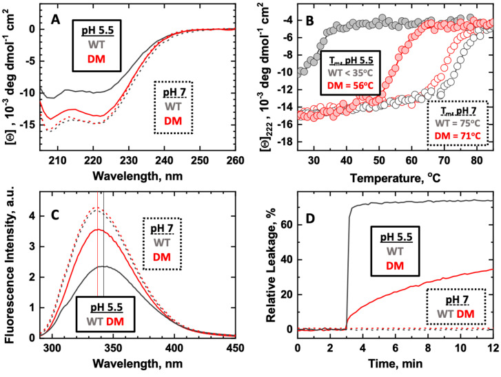
Protonation of key histidine residues has been long implicated in the acid-mediated cellular action of the diphtheria toxin translocation (T-) domain, responsible for the delivery of the catalytic domain into the cell. Here, we use a combination of computational (constant-pH Molecular Dynamics simulations) and experimental (NMR, circular dichroism, and fluorescence spectroscopy along with the X-ray crystallography) approaches to characterize the initial stages of conformational change happening in solution in the wild-type T-domain and in the H223Q/H257Q double mutant. This replacement suppresses the acid-induced transition, resulting in the retention of a more stable protein structure in solutions at pH 5.5 and, consequently, in reduced membrane-disrupting activity. Here, for the first time, we report the pKa values of the histidine residues of the T-domain, measured by NMR-monitored pH titrations. Most peaks in the histidine side chain spectral region are titrated with pKas ranging from 6.2 to 6.8. However, the two most up-field peaks display little change down to pH 6, which is a limiting pH for this protein in solution at concentrations required for NMR. These peaks are absent in the double mutant, suggesting they belong to H223 and H257. The constant-pH simulations indicate that for the T-domain in solution, the pKa values for histidine residues range from 3.0 to 6.5, with those most difficult to protonate being H251 and H257. Taken together, our experimental and computational data demonstrate that previously suggested cooperative protonation of all six histidines in the T-domain does not occur.

Keywords: NMR spectroscopy; X-ray crystallography; acidification; conformational switching; constant-pH simulations; diphtheria toxin structure; mutations of histidines.


Conflict of interest statement

The authors declare no conflict of interest.

Figures

Figure 1
Figure 1
General scheme of acid-induced conformational switching and membrane insertion of Diphtheria Toxin T-domain (for details see [21,22]). The insertion pathway is initiated by the conversion of the water-soluble unprotonated W-state into the protonated membrane-competent state W+-state and its subsequent binding to the membrane interface. The conversion from a family of interfacial states into two predominant transmembrane states, with different topologies of the N-terminus, is facilitated by the presence of anionic lipids. The exact molecular mechanism of the translocation of the Catalytic domain of the toxin, attached to the T-domain’s N-terminus, remains unknown. The evidence from mutagenesis indicates that the formation of the so-called Open-Channel State, OCS (illustrated by the cartoon on the right) is not necessary for the translocation and that the OCS does not constitute the translocation pathway [23] (the six native histidines are listed in the scheme and are highlighted in red. The consensus insertion hairpin formed by helices TH8 and TH9 is highlighted in brown. Helix TH5, which can have either interfacial or transmembrane topology, is highlighted in blue in the two membrane-inserted conformations. No high-resolution structures are available for the T-domain in the lipid bilayer, and the presented schemes, developed from various spectroscopic and computational experiments, are shown for illustration purposes only). The focus of this study is on the initial conformational transition occurring in solution (red ractangle), which is associated with the protonation of the histidine residues.
Figure 2
Figure 2
Comparison of pH-induced conformational switching (AC) and membrane action (D) of the T-domain wild type (WT, black lines) and H223Q/H257Q Double Mutant (DM, red lines). Dashed curves and open symbols correspond to pH 7, and solid curves and symbols to pH 5.5. The replacement of the two histidines with non-protonatable residues suppresses conformational change leading to the formation of the membrane-competent W+-state, as seen by the reduced changes in CD (A) and tryptophan fluorescence spectra (C). The thermal stability of the helical structure, as measured by changes of ellipticity at 222 nm, is much higher at pH 5.5 for the DM than that for the WT T-domain (B). Subsequently, the ability of the DM to induce the leakage of the membrane content at pH 5.5 is much reduced compared to the WT (D).
Figure 3
Figure 3
Comparison of the high-resolution crystallographic structures of the Diphtheria Toxin dimers: WT at pH 7 (A), 5.5 (B), and DM at pH 5.5 (C). In each case, the toxin forms a domain-swapped dimer shown, with one chain colored uniformly in grey and the other colored in blue (Receptor-binding domain), green (Translocation domain), and red (Catalytic domain). The six histidines are shown only for the former chain in panel A and are colored the same way as their corresponding labels. The first two structures, published in our previous study [34], reveal acid-induced loss of the helical structure of TH2 of the WT T-domain (labeled panel A as TH2-A and TH2-B for each monomer). This change is not observed when H223 and H257 are replaced in the DM of both crystal forms (PDB 8G0F and 8G0G) (C). Otherwise, the overall fold is not affected by the replacement of the two histidines (local rearrangements are shown in Figure 4).
Figure 4
Figure 4
The local rearrangement of the packing around residues 223 and 257 in the DM vs. the WT T-domain. Q223 in DM (8G0G, gold/orange) forms a hydrogen bond with E259. This results in the movement of residue Q257 relative to the WT structure (7K7E, magenta/gray), and a new sidechain interaction is formed with T253.
Figure 5
Figure 5
pH-dependent changes in the histidine side chain NMR peaks in the T-domain WT (A) and DM (B). The two WT peaks (panel A) that show no change (Peak A and Peak B) are likely to belong to H223 and H257, as the corresponding peaks are absent in the DM (panel B). The rest of the peaks appear to have very similar patterns in DM and WT (note that the number of the peaks in the region between 8.3 and 7.5 ppm is greater than the number of histidine side chains in both the WT and DM construct. This discrepancy is attributed to either dynamic conformational heterogeneity or overlap from other side chain resonances. Panels C and D represent the support-plane analysis of the titratable peaks in the WT (panel C) and the DM T-domain (panel D), which provides a measure of the reliability of pKa determination. The minimum of the curve corresponds to the most probable pKa, while the intercepts with the solid line at 1.33 corresponds to a range of one standard deviation. Even for the most visually different curves corresponding to the Peak G, the resulting confidence intervals of pKa overlap for the WT ((C), pKa = [6.2–6.5]) and the DM ((D) pKa = [5.8–6.3]).
Figure 6
Figure 6
Summary of the constant-pH MD simulations of the WT T-domain. The individual pH titration curves and calculated pKa values for residues H223, H251, H257, and E259 (A) and H322, H323, and H372 (B). Structure representation of DTT after 150 ns at pH 4.5 (C). The protein is shown in the cartoon (light gray) with TH1 highlighted in blue and TH2 in red color, and with residues H223 (red), H257 (blue), and Glu259 (olive) represented as sticks. The average distances between H257 and E259 at different pH values illustrate the acidic residue sequestering induced by protonation (D). The average helicity content of TH2, at different pH values at three different replicates, highlights the significant loss of structure in one replicate (R3) induced by H257 protonation (E).

References

    1. Hoch D.H., Romero-Mira M., Ehrlich B.E., Finkelstein A., DasGupta B.R., Simpson L.L. Channels formed by botulinum, tetanus, and diphtheria toxins in planar lipid bilayers: Relevance to translocation of proteins across membranes. Proc. Natl. Acad. Sci. USA. 1985;82:1692. doi: 10.1073/pnas.82.6.1692. - DOI - PMC - PubMed
    1. Arnon S.S., Schechter R., Inglesby T.V., Henderson D.A., Bartlett J.G., Ascher M.S., Eitzen E., Fine A.D., Hauer J., Layton M., et al. Botulinum toxin as a biological weapon: Medical and public health management. JAMA. 2001;285:1059–1070. doi: 10.1001/jama.285.8.1059. - DOI - PubMed
    1. Neale E.A. Moving across membranes. Nature Struct. Biol. 2003;10:2–3. doi: 10.1038/nsb0103-2. - DOI - PubMed
    1. Koriazova L.K., Montal M. Translocation of botulinum neurotoxin light chain protease through the heavy chain channel. Nat. Struct. Biol. 2003;10:13–18. doi: 10.1038/nsb879. - DOI - PubMed
    1. Collier R.J., Young J.A. Anthrax toxin. Annu. Rev. Cell Dev. Biol. 2003;19:45–70. doi: 10.1146/annurev.cellbio.19.111301.140655. - DOI - PubMed

Publication types

LinkOut - more resources