

Fecal Calprotectin Concentrations in Cats with Chronic Enteropathies
- PMID: 37505825
- PMCID: PMC10385529
- DOI: 10.3390/vetsci10070419
Fecal Calprotectin Concentrations in Cats with Chronic Enteropathies
Abstract
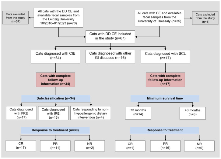
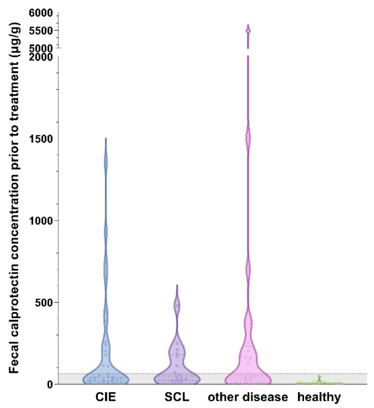
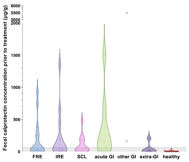
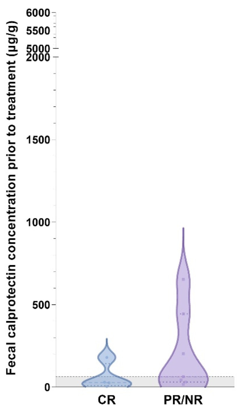
Diagnosis of feline chronic inflammatory enteropathies (CIE) and the differentiation from small cell intestinal lymphoma (SCL) can be challenging. Intestinally expressed calprotectin (S100A8/A9 protein complex) appears to be part of the complex pathogenesis of feline chronic enteropathies (FCE). Fecal calprotectin is a non-invasive biomarker for intestinal inflammation in humans and dogs but has not yet been evaluated in cats. We hypothesized that fecal calprotectin (fCal) concentrations are increased in FCE, correlate with clinical and/or histologic disease severity, and distinguish cases of CIE from SCL. This case-control study included fecal samples and patient data from cats with CIE (n = 34), SCL (n = 17), other gastrointestinal (GI) diseases (n = 16), and cats with no clinical signs of GI disease (n = 32). fCal concentrations were measured using the immunoturbidimetric fCal turbo assay (Bühlmann Laboratories). Compared to healthy cats, fCal concentrations were significantly increased in CIE, SCL, and other diseases (all p < 0.0001), but were not different between these three groups (all p > 0.05), or between cats with extra-GI diseases and healthy controls. These findings suggest that fCal may have utility as a clinical biomarker for FCE but not for intestinal disease differentiation. It further supports the role of calprotectin in the pathogenesis of the spectrum of FCE, which includes CIE and SCL.
Keywords: biomarker; feline chronic enteropathy; food-responsive enteropathy; gastrointestinal diet; hydrolyzed diet; inflammatory bowel disease; small cell lymphoma; steroid-responsive enteropathy.
Conflict of interest statement
Our study was performed with the generous support of Bühlmann Laboratories, Schönenbuch, BL, Switzerland. The company provided analyzer reagents, expertise in the application, and assay-specific materials. The funders had no role in the design of the study; in the collection, analyses, or interpretation of data; in the writing of the manuscript, or in the decision to publish the results.
Figures
Similar articles
-
Intestinal S100/Calgranulin Expression in Cats with Chronic Inflammatory Enteropathy and Intestinal Lymphoma.Animals (Basel). 2022 Aug 11;12(16):2044. doi: 10.3390/ani12162044. Animals (Basel). 2022. PMID: 36009635 Free PMC article.
-
Treatment success in cats with chronic enteropathy is associated with a decrease in fecal calprotectin concentrations.Front Vet Sci. 2024 Apr 3;11:1390681. doi: 10.3389/fvets.2024.1390681. eCollection 2024. Front Vet Sci. 2024. PMID: 38634105 Free PMC article.
-
Verification of the fCAL turbo immunoturbidimetric assay for measurement of the fecal calprotectin concentration in dogs and cats.J Vet Diagn Invest. 2022 Sep;34(5):813-824. doi: 10.1177/10406387221114031. Epub 2022 Jul 25. J Vet Diagn Invest. 2022. PMID: 35879875 Free PMC article.
-
Clinical utility of currently available biomarkers in inflammatory enteropathies of dogs.J Vet Intern Med. 2018 Sep;32(5):1495-1508. doi: 10.1111/jvim.15247. Epub 2018 Sep 17. J Vet Intern Med. 2018. PMID: 30222209 Free PMC article. Review.
-
Differentiating Inflammatory Bowel Disease from Alimentary Lymphoma in Cats: Does It Matter?Vet Clin North Am Small Anim Pract. 2021 Jan;51(1):93-109. doi: 10.1016/j.cvsm.2020.09.009. Vet Clin North Am Small Anim Pract. 2021. PMID: 33187624 Review.
Cited by
-
Effects of Chicken Protein Hydrolysate as a Protein Source to Partially Replace Chicken Meal on Gut Health, Gut Microbial Structure, and Metabolite Composition in Cats.Vet Sci. 2025 Apr 21;12(4):388. doi: 10.3390/vetsci12040388. Vet Sci. 2025. PMID: 40284890 Free PMC article.
-
Plasma glucagon-like peptide-2 in cats with chronic enteropathies.J Feline Med Surg. 2025 Jan;27(1):1098612X241305923. doi: 10.1177/1098612X241305923. J Feline Med Surg. 2025. PMID: 39840661 Free PMC article.
-
Effect of Iron Deficiency on Short-Term Response to Treatment in Cats With Chronic Enteropathies.J Vet Intern Med. 2025 May-Jun;39(3):e70131. doi: 10.1111/jvim.70131. J Vet Intern Med. 2025. PMID: 40384256 Free PMC article.
-
Duodenal and colonic mucosal S100A8/A9 (calprotectin) expression is increased and correlates with the severity of select histologic lesions in dogs with chronic inflammatory enteropathy.BMC Vet Res. 2024 Sep 6;20(1):393. doi: 10.1186/s12917-024-04256-9. BMC Vet Res. 2024. PMID: 39238011 Free PMC article.
-
Evaluation of the fatty acid-based erythrocyte membrane lipidome in cats with food responsive enteropathy, inflammatory bowel disease and low-grade intestinal T-cell lymphoma.PLoS One. 2024 Jul 29;19(7):e0307757. doi: 10.1371/journal.pone.0307757. eCollection 2024. PLoS One. 2024. PMID: 39074116 Free PMC article.
References
-
- Washabau R.J., Day M.J., Willard M.D., Hall E.J., Jergens A.E., Mansell J., Minami T., Bilzer T.W. Endoscopic, Biopsy, and Histopathologic Guidelines for the Evaluation of Gastrointestinal Inflammation in Companion Animals. J. Vet. Intern. Med. 2010;24:10–26. doi: 10.1111/J.1939-1676.2009.0443.X. - DOI - PubMed
Grants and funding
LinkOut - more resources
Full Text Sources
Research Materials
Miscellaneous
