

Development and Application of nanoPCR Method for Detection of Feline Panleukopenia Virus
- PMID: 37505845
- PMCID: PMC10386105
- DOI: 10.3390/vetsci10070440
Development and Application of nanoPCR Method for Detection of Feline Panleukopenia Virus
Abstract
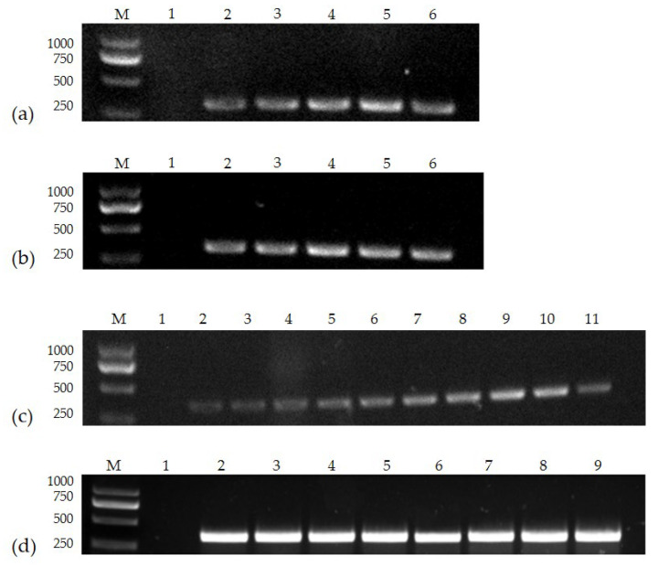
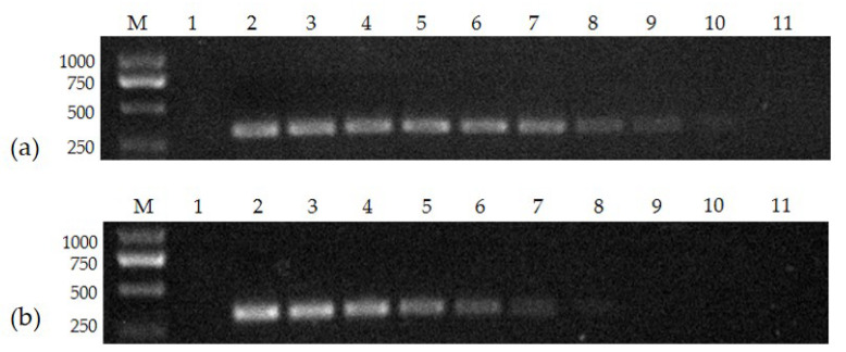
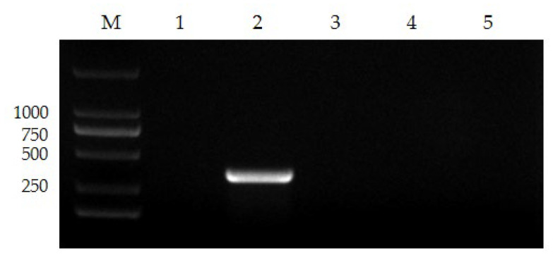
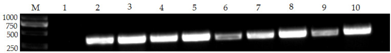
Feline panleukopenia (FP) is a severe viral illness caused by the feline panleukopenia virus (FPV), putting sectors like companion cat breeding and endangered feline conservation at risk. The virus has a high morbidity and fatality rate and is found all over the world. We created a novel FPV assay using nanoPCR technology and assessed the method's specificity and sensitivity. The approach amplified a 345 bp nucleic acid fragment with a minimum detection limit of 7.97 × 10² copies/μL, which is about 100 times greater than traditional PCR. We collected anal swabs from 83 cats suspected of FPV infection for practical application, and the FPV-positive rate determined by the nanoPCR approach was 77.1%. In conclusion, the approach is more sensitive than conventional PCR and more convenient and cost-effective than qPCR methodology and may be utilized for the clinical detection of FPV.
Keywords: VP2 gene; clinical testing; epidemiology; feline panleukopenia virus; nanoPCR.
Conflict of interest statement
The authors declare no conflict of interest.
Figures
Similar articles
-
Development and Application of an RPA-Based Rapid Point-of-Care Testing (POCT) Method for the Detection of Feline Panleukopenia Virus.Transbound Emerg Dis. 2024 Aug 24;2024:3680778. doi: 10.1155/2024/3680778. eCollection 2024. Transbound Emerg Dis. 2024. PMID: 40303162 Free PMC article.
-
Development of a triple NanoPCR method for feline calicivirus, feline panleukopenia syndrome virus, and feline herpesvirus type I virus.BMC Vet Res. 2022 Oct 27;18(1):379. doi: 10.1186/s12917-022-03460-9. BMC Vet Res. 2022. PMID: 36303189 Free PMC article.
-
Feline panleukopenia virus DNA shedding following modified live virus vaccination in a shelter setting.Vet J. 2022 Jan;279:105783. doi: 10.1016/j.tvjl.2021.105783. Epub 2021 Nov 30. Vet J. 2022. PMID: 34861370
-
Feline parvovirus infection and associated diseases.Vet J. 2014 Aug;201(2):150-5. doi: 10.1016/j.tvjl.2014.05.027. Epub 2014 May 22. Vet J. 2014. PMID: 24923754 Review.
-
Feline panleukopenia virus: its interesting evolution and current problems in immunoprophylaxis against a serious pathogen.Vet Microbiol. 2013 Jul 26;165(1-2):29-32. doi: 10.1016/j.vetmic.2013.02.005. Epub 2013 Feb 18. Vet Microbiol. 2013. PMID: 23561891 Review.
Cited by
-
Development and Application of an RPA-Based Rapid Point-of-Care Testing (POCT) Method for the Detection of Feline Panleukopenia Virus.Transbound Emerg Dis. 2024 Aug 24;2024:3680778. doi: 10.1155/2024/3680778. eCollection 2024. Transbound Emerg Dis. 2024. PMID: 40303162 Free PMC article.
-
The quadruplex TaqMan MGB fluorescent quantitative PCR method for simultaneous detection of feline panleukopenia virus, feline herpesvirus 1, feline calicivirus and feline infectious peritonitis virus.Front Cell Infect Microbiol. 2025 May 30;15:1581946. doi: 10.3389/fcimb.2025.1581946. eCollection 2025. Front Cell Infect Microbiol. 2025. PMID: 40521026 Free PMC article.
-
Nanomaterials in PCR: exploring light-to-heat conversion mechanisms and microfluidic integration.Microsyst Nanoeng. 2025 Jun 19;11(1):127. doi: 10.1038/s41378-025-00898-3. Microsyst Nanoeng. 2025. PMID: 40533488 Free PMC article. Review.
References
Grants and funding
LinkOut - more resources
Full Text Sources
Miscellaneous
