. 2023 Aug 29;42(8):112859.
doi: 10.1016/j.celrep.2023.112859. Epub 2023 Jul 27.

Condensate cooperativity underlies transgenerational gene silencing

Affiliations

Condensate cooperativity underlies transgenerational gene silencing

Zhenzhen Du et al. Cell Rep. .

Abstract

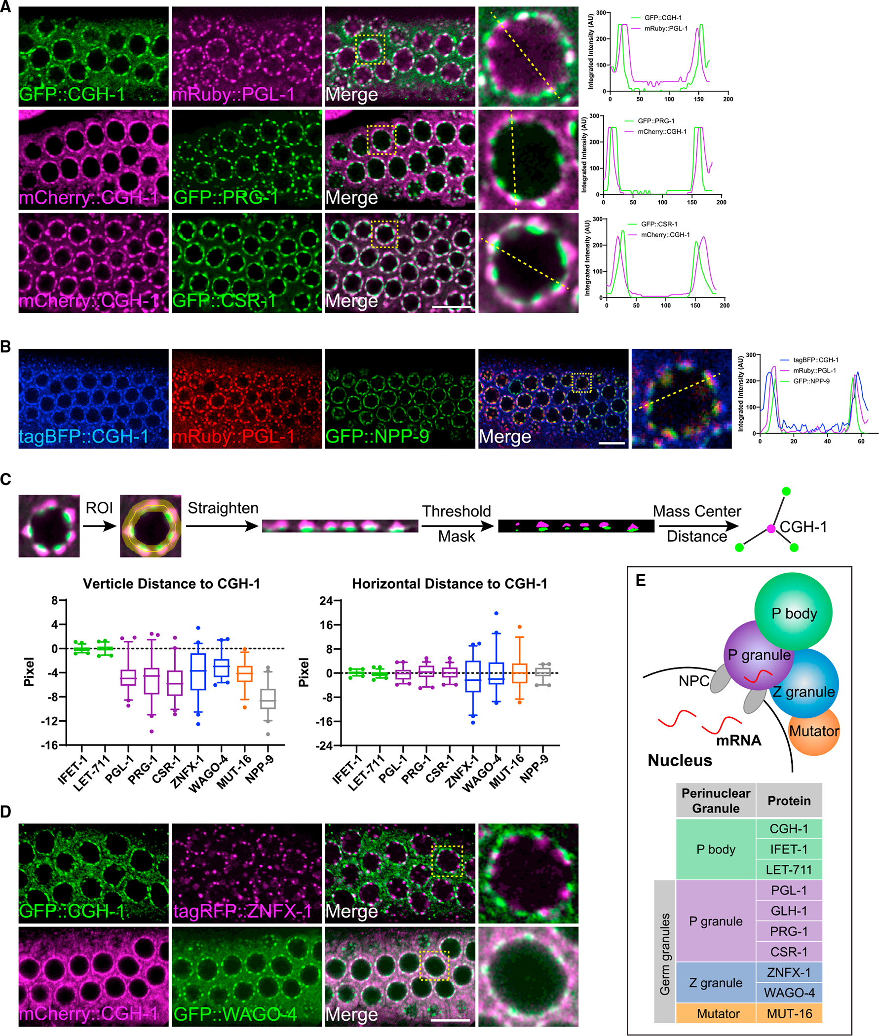
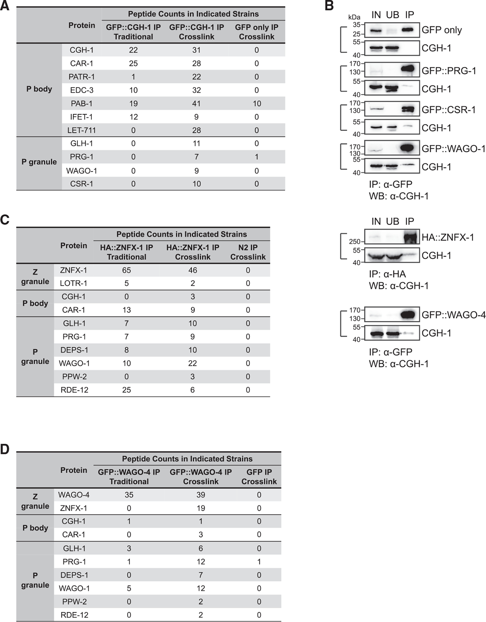
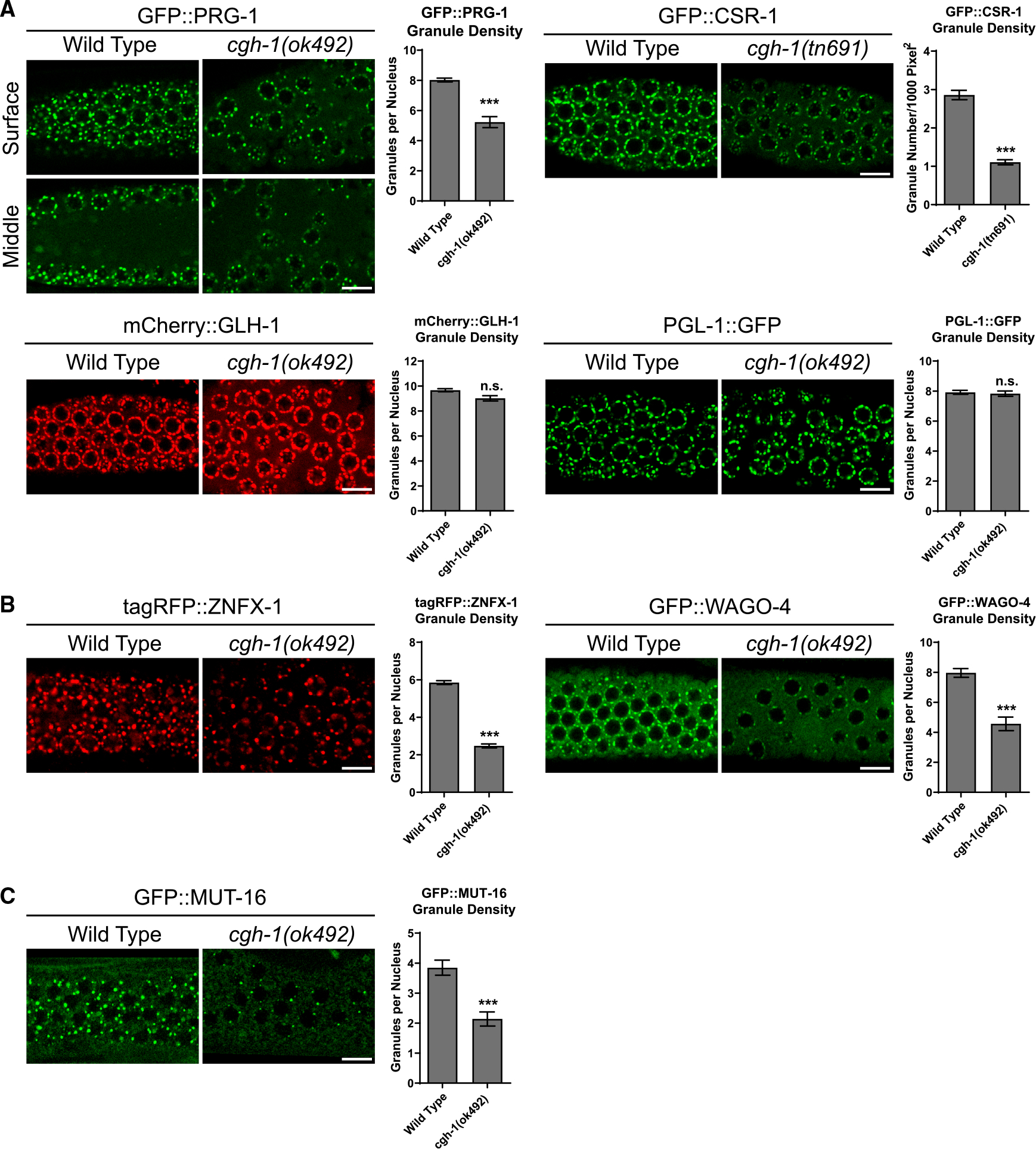
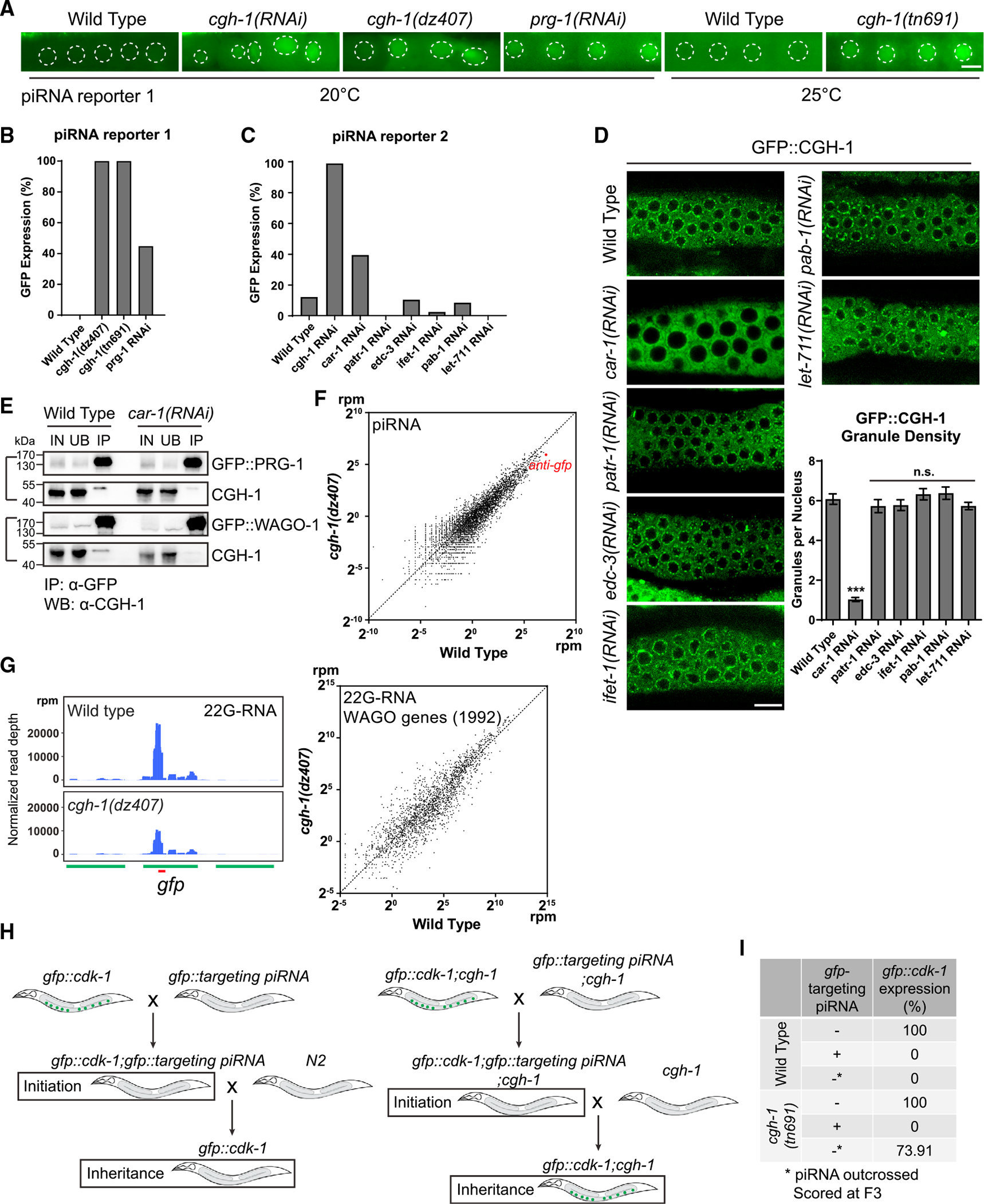
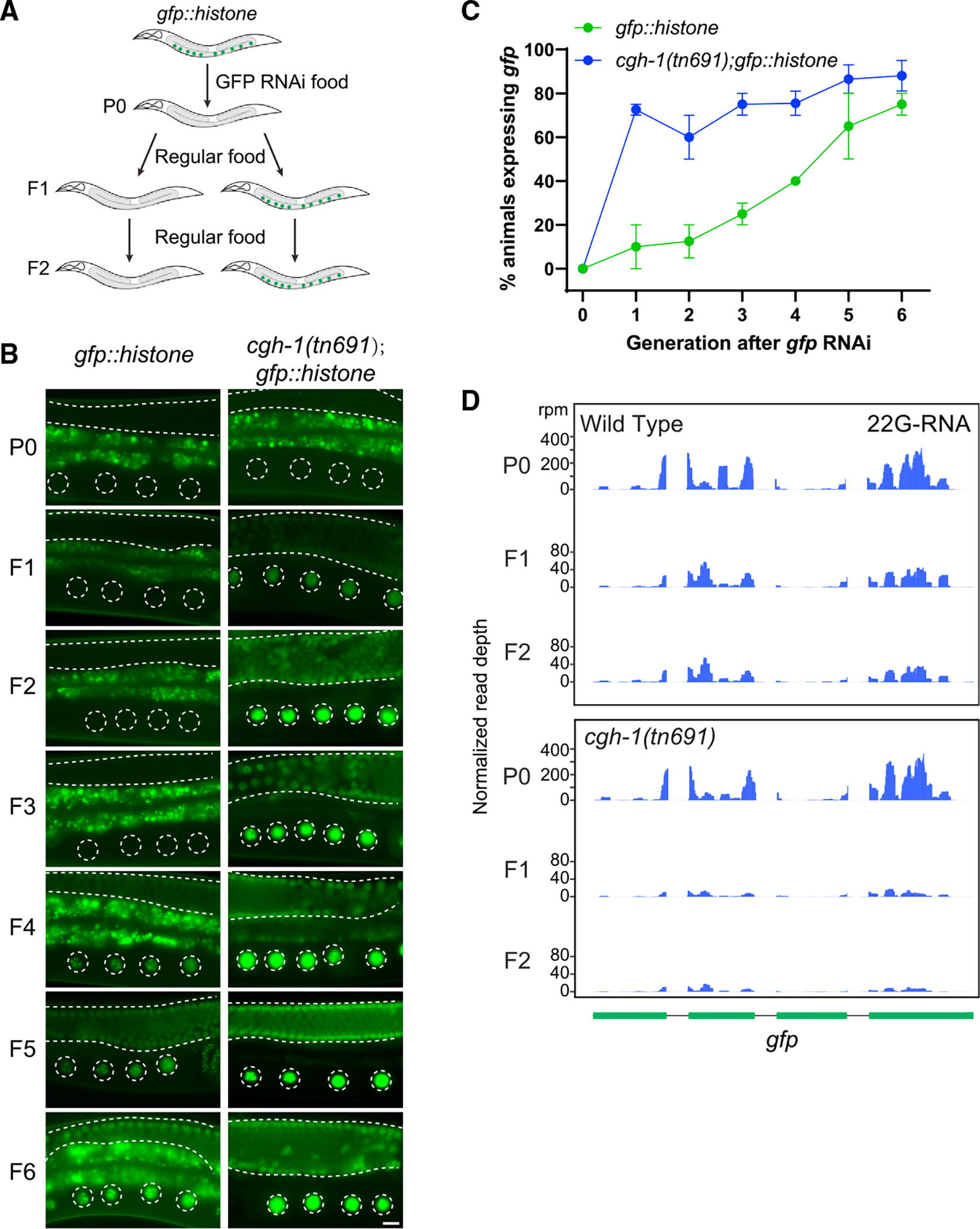
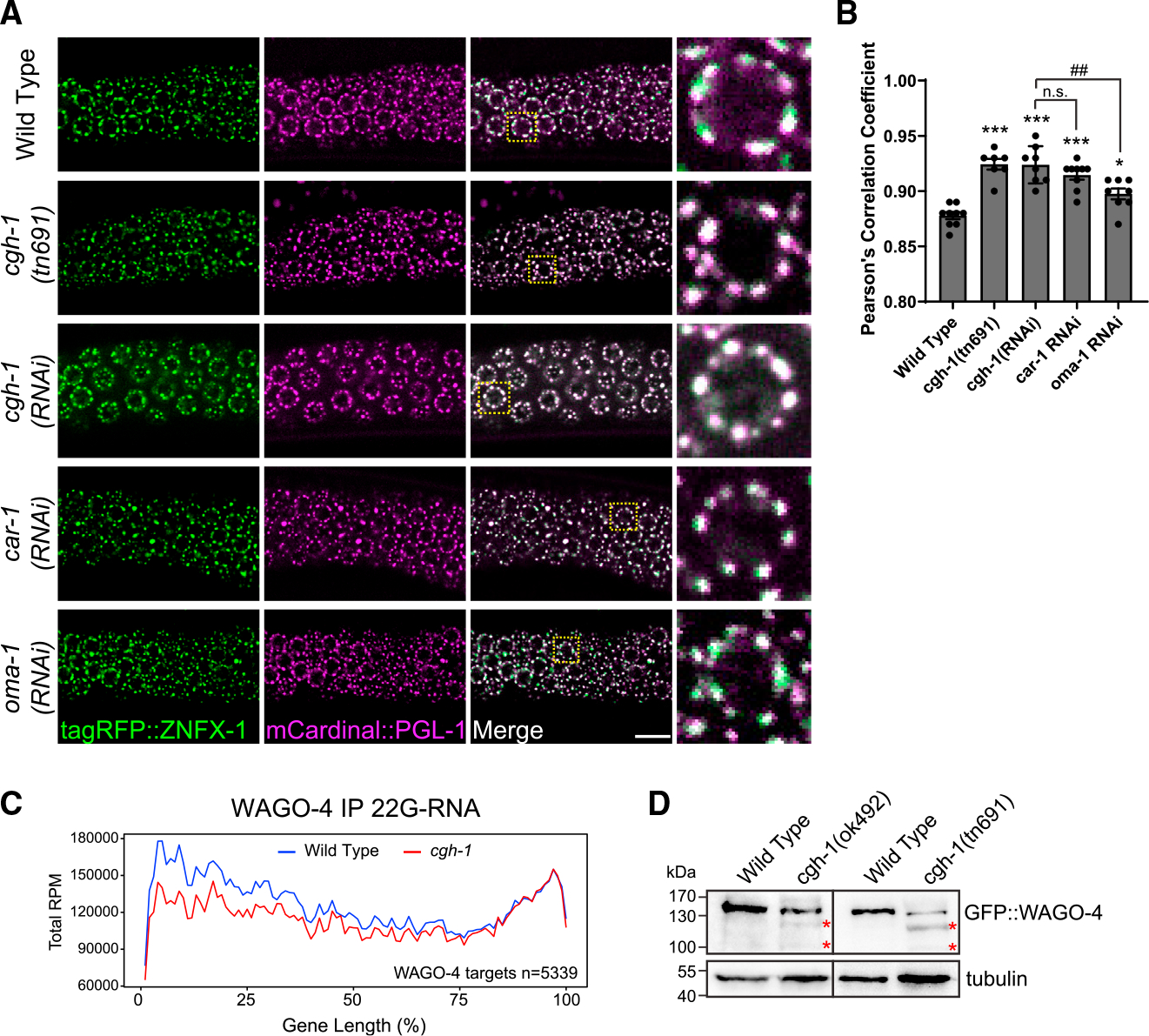
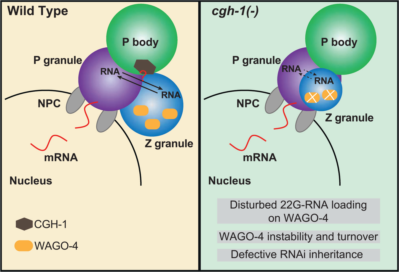
Biomolecular condensates have been shown to interact in vivo, yet it is unclear whether these interactions are functionally meaningful. Here, we demonstrate that cooperativity between two distinct condensates-germ granules and P bodies-is required for transgenerational gene silencing in C. elegans. We find that P bodies form a coating around perinuclear germ granules and that P body components CGH-1/DDX6 and CAR-1/LSM14 are required for germ granules to organize into sub-compartments and concentrate small RNA silencing factors. Functionally, while the P body mutant cgh-1 is competent to initially trigger gene silencing, it is unable to propagate the silencing to subsequent generations. Mechanistically, we trace this loss of transgenerational silencing to defects in amplifying secondary small RNAs and the stability of WAGO-4 Argonaute, both known carriers of gene silencing memories. Together, these data reveal that cooperation between condensates results in an emergent capability of germ cells to establish heritable memory.

Keywords: Argonaute; CGH-1; CP: Genomics; CP: Molecular biology; P body; PIWI; condensate; germ cells; germ granule; phase separation; small RNA; transgenerational inheritance.


Conflict of interest statement

Declaration of interests The authors declare no competing interests.

Figures

Figure 1.
Figure 1.. P bodies localize to the cytoplasmic side of perinuclear germ granules
(A) Fluorescent micrographs show the localization of P body marker CGH-1 and the indicated P granule markers in the pachytene region of adult germlines. The line in the merged image indicates the position of the line scan for measuring fluorescent intensity across single germline nuclei (right). Note that white-colored foci in the merged image represent the co-localization of two makers. Bar: 10 μm. (B) Fluorescent micrographs show the spatial arrangement of P body, P granule, and nuclear pore clusters at germline nuclei. The line in the merged image indicates the position of the line scan for measuring fluorescent intensity across single germline nuclei (right). Bar: 10 μm. (C) A schematic (top) showing the measurements of vertical distance (bottom left) and horizontal (bottom right) distance between the indicated proteins to P body marker CGH-1. For boxplots, lines display median values, boxes display 1st and 3rd quartiles, and whiskers display 5th and 95th percentiles. Distributions represent data collected from 8 to 12 independent gonad images. (D) Fluorescent micrographs show the localization of P body marker CGH-1 and the indicated Z granule proteins in the pachytene region of adult germlines. Note that white-colored foci in the merged image represent the co-localization of two markers. Bar: 10 μm. (E) A model depicting spatial arrangements of the P body and distinct sub-compartments of the germ granule, including P granule, Z granule, and Mutator foci, at the nuclear periphery.
Figure 2.
Figure 2.. Proteomic analyses of interactions between P body and germ granule factors
(A) Proteomic analyses of CGH-1 complex. The numbers of peptides identified in the crosslinked or non-crosslinked condition are shown. GFP-only IP experiment in the crosslink condition served as a control. (B) Western blot analyses show the interactions between CGH-1 and the indicated germ granule factors. Immunoprecipitation of GFP- or HA-tagged germ granule factors was conducted. Subsequently, western blot analysis was performed using specific antibodies against GFP, HA, or CGH-1 to determine the presence of the indicated germ granule factors or CGH-1 in the indicated samples. IN, input; UB, unbound fraction; IP, immunoprecipitated fraction. (C) Proteomic analyses of ZNFX-1 complex. The numbers of peptides identified in the crosslinked or non-crosslinked condition are shown. N2 control IP experiment in the crosslink condition served as a control. (D) Proteomic analyses of WAGO-4 complex. The numbers of peptides identified in the crosslinked or non-crosslinked condition are shown. N2 control IP experiment in the crosslink condition served as a control.
Figure 3.
Figure 3.. P body factor CGH-1 promotes the localization of small RNA pathway factors at perinuclear germ granules
Representative fluorescent micrographs and corresponding quantification of granule density of indicated P granule markers (A), Z granule markers (B), and Mutator foci marker MUT-16 (C) in the pachytene region of adult germlines in wild-type or in cgh-1 (ok492) mutant animals. Bars: 10 μm. The images were captured from the surface section of the germline. Additionally, images of the middle section of the germline of GFP::PRG-1 were included to highlight the observed defects in germline nuclei organization in cgh-1 (ok492) mutant. Statistical analysis was performed using a one-tailed Student’s t test. Bars indicate the mean, and errors bars indicate the standard deviation. Distributions represent data collected from 8 to 12 independent gonad images.
Figure 4.
Figure 4.. P body factors CGH-1 and CAR-1 promote piRNA-dependent gene silencing
(A) GFP expression of the piRNA reporter #1 in the indicated strains. In this reporter, the expressions of both CDK-1:GFP (nuclear) and OMA-1:GFP (cytoplasm) are silenced by a synthetic GFP-targeting piRNA in wild-type animals. Worms were cultured at 20°C for imaging. For cgh-1 (tn691) mutant and its control wild-type animals, worms were grown till young adult stage at 20° and subsequently transferred to 25°C for a 12-h period before imaging. Dotted circles indicate the location of maturing oocyte nuclei. Bars: 10 μm. (B) Percentage of screened animals showing GFP expression in the piRNA reporter #1 in the indicated strains. We define the expression of GFP (on/off) by observing whether the indicated mutant or RNAi-treated animals exhibit visible nuclear and cytoplasmic GFP signals in their germline. (C) Percentage of screened animals showing GFP expression in the piRNA reporter #2 in the indicated strains. We define the expression of GFP (on/off) by observing whether the indicated RNAi-treated animals exhibit visible nuclear and cytoplasmic GFP signals in their germline. (D) Fluorescent micrographs show the localization of P body marker CGH-1 in the indicated RNAi-treated animals. Bars: 10 μm. Granule density (number of granule/nuclei/section) of GFP::CGH-1 in wild-type or the indicated RNAi-treated animals was calculated. These quantifications correspond to micrographs in Figure 4D. Statistical analysis was performed using a one-tailed Student’s t test. Bars indicate the mean, and errors bars indicate the standard deviation. Distributions represent data collected from 8 to 12 independent gonad images. (E) Western blot analyses showing that the interaction between CGH-1 and P granule factors PRG-1 or WAGO-1 is reduced in car-1 RNAi-treated animals. IN, input; UB, unbound fraction; IP, immunoprecipitated fraction. (F) A scatterplot showing the abundance of piRNAs in wild-type worms compared to cgh-1 (dz407) mutant. The red dot indicates the levels of the synthetic GFP-targeting piRNA. (G) 22G-RNAs distribution at GFP coding sequences in the indicated strains. The red bar indicates the location of the GFP sequence complementary to the GFP-targeting piRNA (left). A scatterplot shows the abundance of 22G-RNAs mapped to WAGO target genes in wild-type worms compared to cgh-1 (dz407) mutant (right). (H) A schematic showing the GFP reporter assays to determine the role of CGH-1 in the initiation or maintenance of piRNA silencing. (I) Percentage of GFP reporter expression from screened animals in the indicated strains. Note that cgh-1(tn691) mutant animals can initiate piRNA-directed silencing of the GFP reporter but exhibit defects in the inheritance of gene silencing.
Figure 5.
Figure 5.. CGH-1 promotes RNAi inheritance and 22G-RNA synthesis in inheriting generations
(A) A schematic showing the RNAi assay with a GFP::histone reporter to determine whether the strain can initiate RNAi and inherit gene silencing over generations. In these experiments, both wild-type and cgh-1 (tn691) mutant animals are grown at the permissive temperature (20°C). (B) Fluorescent micrographs show representative images of the GFP::H2B reporter in the RNAi-treated generation (P0) or the following generations (F1 to F6) in wild-type and cgh-1 (tn691) mutant animals. Dashed circles indicate the location of maturing oocytes nuclei, and the area between two dashed lines indicates germline tissue. Note that the bright signals observed outside of these two highlighted areas are autofluorescence signals originating from C. elegans gut granules. Bars: 10 μm. (C) Percentage of screened animals showing GFP reporter expression in wild-type and cgh-1 (tn691) mutant animals. Statistical analysis was performed using a one-tailed Student’s t test. Data points indicate the mean, and errors bars indicate the standard deviation. Distributions represent data collected from three independent experiments. (D) Anti-sense 22G-RNAs distribution mapped to GFP coding sequences in the indicated strains from the RNAi-treated animals (P0) or the following generations (F1 or F2).
Figure 6.
Figure 6.. CGH-1 promotes proper germ granule organization and WAGO-4 22G-RNA production
(A) Fluorescent micrographs show the localization of P granule marker PGL-1 and Z granule marker ZNFX-1 in the indicated mutant or RNAi-treated animals. Bars: 10 μm. (B) Pearson’s correlation coefficient of PGL-1 and ZNFX-1 signals in the indicated mutant or RNAi-treated animals. Bars indicate the mean, and errors bars indicate the standard deviation. Distributions represent data collected from 8 to 12 independent gonad images. (C) Metagene traces show the total accumulation of WAGO-4-associated 22G-RNAs by percentage of WAGO-4 target gene length in IP experiments. Reads from wild-type IP experiments are traced in blue, and reads from cgh-1 RNAi-treated IP experiments are traced in red. (D) Western blots show the levels of WAGO-4 in wild type or in the indicated cgh-1 mutants. The red asterisks indicated partially degraded WAGO-4 proteins.
Figure 7.
Figure 7.. A model showing the roles of P body factor CGH-1 in germ granule organization and WAGO-4 22G-RNA production that promote transgenerational gene silencing

Similar articles

Cited by

References

    1. Banani SF, Lee HO, Hyman AA, and Rosen MK (2017). Biomolecular condensates: organizers of cellular biochemistry. Nat. Rev. Mol. Cell Biol. 18, 285–298. 10.1038/nrm.2017.7. - DOI - PMC - PubMed
    1. Lyon AS, Peeples WB, and Rosen MK (2021). A framework for understanding the functions of biomolecular condensates across scales. Nat. Rev. Mol. Cell Biol. 22, 215–235. 10.1038/s41580-020-00303-z. - DOI - PMC - PubMed
    1. Fare CM, Villani A, Drake LE, and Shorter J (2021). Higher-order organization of biomolecular condensates. Open Biol. 11, 210137. 10.1098/rsob.210137. - DOI - PMC - PubMed
    1. Claycomb JM, Batista PJ, Pang KM, Gu W, Vasale JJ, van Wolfswinkel JC, Chaves DA, Shirayama M, Mitani S, Ketting RF, et al. (2009). The Argonaute CSR-1 and Its 22G-RNA Cofactors Are Required for Holocentric Chromosome Segregation. Cell 139, 123–134. 10.1016/j.cell.2009.09.014. - DOI - PMC - PubMed
    1. Gu W, Shirayama M, Conte D, Vasale J, Batista PJ, Claycomb JM, Moresco JJ, Youngman EM, Keys J, Stoltz MJ, et al. (2009). Distinct Argonaute-Mediated 22G-RNA Pathways Direct Genome Surveillance in the C. elegans Germline. Mol. Cell. 36, 231–244. 10.1016/j.molcel.2009.09.020. - DOI - PMC - PubMed

Publication types

MeSH terms

Substances