. 2023 Oct 30;35(11):4020-4045.
doi: 10.1093/plcell/koad210.

Deciphering the regulatory network of the NAC transcription factor FvRIF, a key regulator of strawberry (Fragaria vesca) fruit ripening

Affiliations

Deciphering the regulatory network of the NAC transcription factor FvRIF, a key regulator of strawberry (Fragaria vesca) fruit ripening

Xiaojing Li et al. Plant Cell. .

Abstract

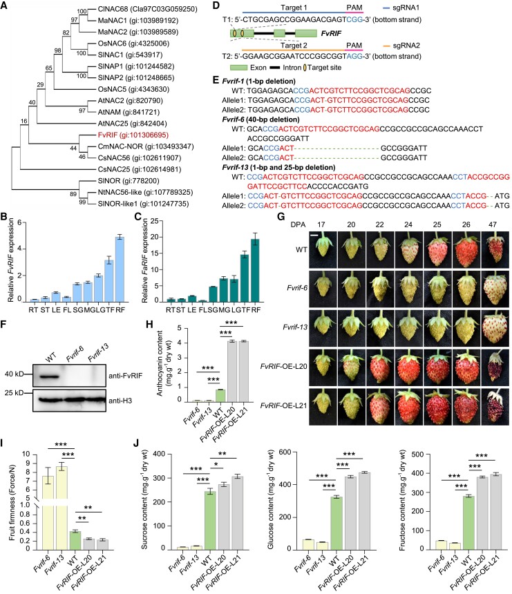
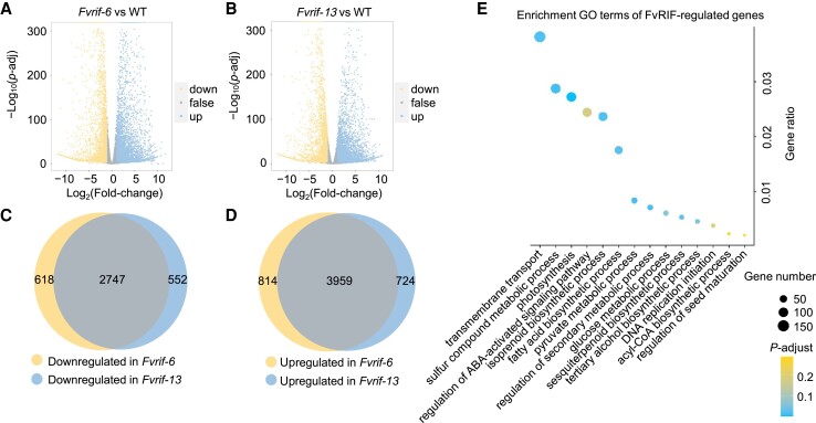
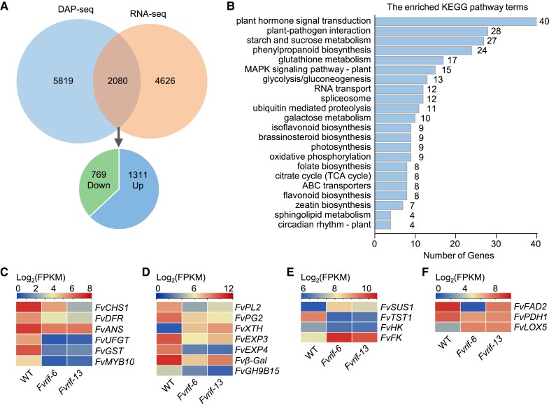
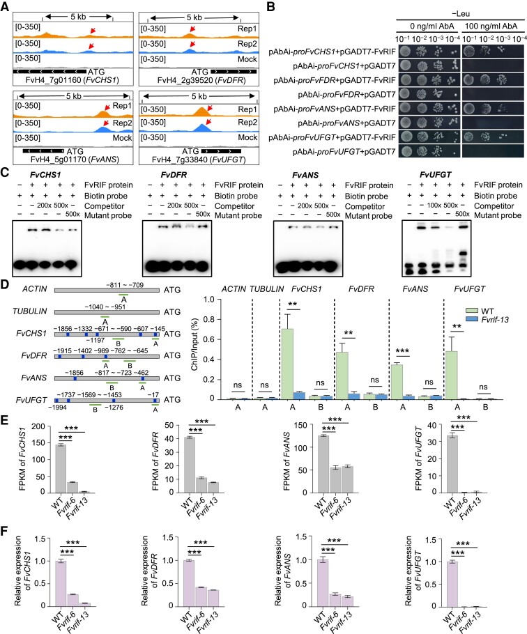
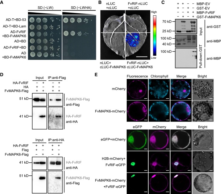
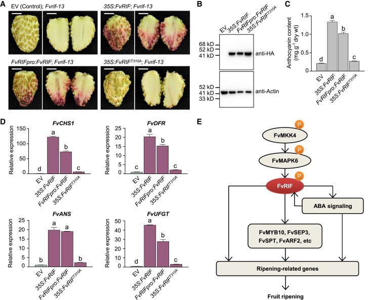
The NAC transcription factor ripening inducing factor (RIF) was previously reported to be necessary for the ripening of octoploid strawberry (Fragaria × ananassa) fruit, but the mechanistic basis of RIF-mediated transcriptional regulation and how RIF activity is modulated remains elusive. Here, we show that FvRIF in diploid strawberry, Fragaria vesca, is a key regulator in the control of fruit ripening and that knockout mutations of FvRIF result in a complete block of fruit ripening. DNA affinity purification sequencing coupled with transcriptome deep sequencing suggests that 2,080 genes are direct targets of FvRIF-mediated regulation, including those related to various aspects of fruit ripening. We provide evidence that FvRIF modulates anthocyanin biosynthesis and fruit softening by directly regulating the related core genes. Moreover, we demonstrate that FvRIF interacts with and serves as a substrate of MAP kinase 6 (FvMAPK6), which regulates the transcriptional activation function of FvRIF by phosphorylating FvRIF at Thr-310. Our findings uncover the FvRIF-mediated transcriptional regulatory network in controlling strawberry fruit ripening and highlight the physiological significance of phosphorylation modification on FvRIF activity in ripening.


Conflict of interest statement

Conflict of interest statement. The authors declare that there is no conflict of interest.

Figures

Figure 1.
Figure 1.
Mutation of FvRIF leads to the inhibition of strawberry fruit ripening. A) Phylogenetic analysis of FvRIF. The phylogenetic tree was generated by MEGA (version 10.1.8). Bootstrap values from 500 replicates for each branch are shown. At, A. thaliana; Cl, C. lanatus; Cm, C. melo; Cs, C. sinensis; Fv, F. vesca; Ma, M. acuminata; Nt, N. tabacum; Os, Oryza sativa; Sl, S. lycopersicum. B), C) Expression pattern of RIF in diploid strawberry (FvRIF, B) and octoploid strawberry (FaRIF, C) as determined by RT-qPCR. FL, flower; LE, leaf; RT, root; ST, stem. Values are means ± standard error of mean (SEM) from 3 biological replicates using ACTIN (FvH4_6g22300) as an internal control. D) Diagram of the sgRNAs with different target sequences (T1 and T2) designed to specifically target exon 1 of FvRIF. E) Genotyping of mutations mediated by CRISPR/Cas9 gene editing in the Fvrif-1, Fvrif-6, and Fvrif-13 mutant lines. Red letters, sgRNA targets; Green dashed lines, edited sites; Blue letters, protospacer adjacent motif (PAM). The transgenic plants in the second generation were genotyped by Sanger sequencing of the genomic regions flanking the target sites. F) Absence of FvRIF protein in the Fvrif-6 and Fvrif-13 lines. Nuclear proteins were extracted from wild-type (WT) and mutant fruits at 28 DPA and subjected to immunoblot analysis using an anti-FvRIF antibody. Histone H3 was used as the internal control. G) Phenotypes of strawberry fruits from WT, Fvrif mutants or FvRIF overexpression lines. Scale bar, 0.5 cm. H) to J) Changes in anthocyanin contents H), fruit firmness I), and sugar contents J) in the mutants and overexpression lines at 28 DPA. Values are means ± SEM from 3 biological replicates. Statistical significance was determined by Student's t-test (*P < 0.05, **P < 0.01, ***P < 0.001).
Figure 2.
Figure 2.
Genome-wide identification of FvRIF binding sites through DAP-seq. A) DAP-seq using 2 biological replicates reveals 9,902 high-confidence FvRIF binding peaks. B) Metaplot of FvRIF binding sites. The FvRIF binding sites are centered on the TSS. TES, transcription end site. C) Distribution of FvRIF binding peaks across genomic features. D) DNA logos of enriched DNA binding sites for FvRIF as determined by Homer (version 3). The P-values are shown. E) Top enriched GO terms of FvRIF-bound genes determined by DAP-seq. Bigger dots indicate more genes.
Figure 3.
Figure 3.
Transcriptome-based determination of FvRIF-regulated genes. A), B) Volcano plots revealing the DEGs in fruits of Fvrif-6A) and Fvrif-13B) mutant lines compared to wild type (WT) at 28 DPA by RNA-seq. Down, downregulated; Up, upregulated. Genes with more than two-fold changes in expression and FDR < 0.05 were considered as DEGs. C), D) Venn diagrams showing the overlapping downregulated C) and upregulated D) genes, respectively, in fruits of the 2 mutant lines (Fvrif-6 and Fvrif-13). E) Top enriched GO terms of FvRIF-regulated genes as defined by RNA-seq analysis. Bigger dots indicate more gene numbers.
Figure 4.
Figure 4.
Identification of direct FvRIF-regulated target genes. A) Venn diagram showing the overlap between FvRIF-bound genes as revealed by DAP-seq and FvRIF-regulated genes identified by RNA-seq. The numbers of downregulated and upregulated FvRIF direct genes are indicated. B) Top KEGG-enriched terms for the direct FvRIF-regulated target genes. C) to F) Heatmap showing the expression of direct FvRIF-regulated target genes related to anthocyanin biosynthesis C), cell-wall degradation D), sugar metabolism E), and aroma compounds generation F). The expression of these genes revealed by RNA-seq in fruits of the mutant lines (Fvrif-6 and Fvrif-13) and the wild type (WT) is shown. The detailed information is shown in Supplemental Data Set S7.
Figure 5.
Figure 5.
FvRIF-mediated direct regulation of genes responsible for anthocyanin biosynthesis in strawberry. A) FvRIF binding peaks (Repeats 1 and 2) and negative control (mock) over the FvCHS1, FvDFR, FvANS, and FvUFGT loci as determined by DAP-seq. [0 to 350] represents the scale of binding intensity as reflected by the height of the peak. UFGT, UDP-glucose flavonoid glucosyl-transferase. B) Y1H assay revealing the binding of FvRIF to the indicated promoter fragments. pGADT7-FvRIF served as the prey and pAbAi-proFvCHS1, proFvDFR, proFvANS, and proFvUFGT were used as the baits. The empty pGADT7 served as the negative control. The transformants were selected on SD/–Leu medium with AbA. C) EMSA shows that FvRIF directly binds to sequence motifs in the FvCHS1, FvDFR, FvANS, and FvUFGT promoters. Recombinant purified FvRIF (0.8 µg) was incubated with biotin-labeled probes (0.02 µm) or unlabeled DNA probe with intact (competitor) or mutated (mutant probe) FvRIF binding motifs. D) ChIP-qPCR assays reveal the direct binding of FvRIF to the promoters of the indicated genes. The promoter structures of the target genes are shown (left panel). Blue boxes, FvRIF binding motifs as identified by DAP-seq; Green lines, regions used for ChIP-qPCR. The numbers indicate the positions of these motifs relative to the ATG. Values are the percentage of DNA fragments coimmunoprecipitated with anti-FvRIF antibodies in fruits of wild type (WT) or the Fvrif-13 mutant line relative to the input DNAs (right panel). Negative controls (ACTIN and TUBULIN) are included. E) Expression of the indicated genes in fruits of Fvrif lines (Fvrif-6 and Fvrif-13) and WT at 28 DPA by RNA-seq. F) Verification of gene expression by RT-qPCR. ACTIN was used as an internal control. For D) to F), values are means ± SEM from 3 biological replicates. Asterisks indicate significant differences (**P < 0.01, ***P < 0.001; Student's t-test).
Figure 6.
Figure 6.
FvRIF-mediated direct regulation of genes involved in fruit softening. A) FvRIF binding peaks (Repeats 1 and 2) and negative control (mock) over the FvPL2, FvPG2, FvXTH, and FvEXP3 loci as determined by DAP-seq. [0 to 350] represents the intensity of binding as reflected by the height of the peak. B) Y1H assay revealing the binding of FvRIF to the indicated promoter fragments. pGADT7-FvRIF served as the prey and the pAbAi-proFvPL2, proFvPG2, proFvXTH, and proFvEXP3 were used as the baits. Empty pGADT7 served as the negative control. The transformants were selected on SD/–Leu medium with AbA. C) EMSA shows that FvRIF directly binds to sequence motifs in the FvPL2, FvPG2, FvXTH, and FvEXP3 promoters. Recombinant purified FvRIF (0.8 µg) was incubated with biotin-labeled probes (0.02 µm) or unlabeled DNA probe with intact (competitor) or mutated (mutant probe) FvRIF binding motifs. D) ChIP-qPCR assays revealing the direct binding of FvRIF to the promoters of the indicated genes. The promoter structures of the target genes are shown (left panel). Blue boxes, FvRIF binding motifs as identified by DAP-seq; Green lines, regions used for ChIP-qPCR. The numbers indicate the positions of these motifs relative to the ATG. Values are the percentage of DNA fragments coimmunoprecipitated with anti-FvRIF antibodies in fruits of wild-type (WT) or the Fvrif-13 mutant line relative to the input DNAs (right panel). Negative controls (ACTIN and TUBULIN) are included. E) Expression of the indicated genes in fruits of Fvrif lines (Fvrif-6 and Fvrif-13) and WT at 28 DPA by RNA-seq. F) Verification of gene expression by RT-qPCR. ACTIN was used as an internal control. For D) to F), values are means ± SEM from 3 biological replicates. Asterisks indicate significant differences (***P < 0.001; Student's t-test).
Figure 7.
Figure 7.
FvRIF-mediated direct regulation of TF genes involved in fruit ripening. A) FvRIF binding peaks (Repeats 1 and 2) and negative control (mock) over the FvMYB10, FvSEP3, FvSPT, and FvARF2 loci as determined by DAP-seq. [0 to 350] represents the intensity of binding as reflected by the heights of the peak. B) Y1H assay revealing the binding of FvRIF to the indicated promoter fragments. pGADT7-FvRIF served as the prey and pAbAi-proFvMYB10, proFvSEP3, proFvSPT, and proFvARF2 were used as the baits. Empty pGADT7 served as the negative control. The transformants were selected on SD/–Leu medium with AbA. C) EMSA shows that FvRIF directly binds to the motifs in the FvMYB10, FvSEP3, FvSPT, and FvARF2 promoters. Recombinant purified FvRIF (0.8 µg) was incubated with biotin-labeled probes (0.02 µm) or unlabeled DNA probe with intact (competitor) or mutated (mutant probe) FvRIF binding motifs. D) ChIP-qPCR assays reveal the direct binding of FvRIF to the promoters of the indicated genes. The promoter structures of the target genes are shown (left panel). Blue boxes, FvRIF binding motifs as identified by DAP-seq; Green lines, regions used for ChIP-qPCR. The numbers indicate the positions of these motifs relative to the ATG. Values are the percentage of DNA fragments coimmunoprecipitated with anti-FvRIF antibodies in fruits of wild-type (WT) or the Fvrif-13 mutant line relative to the input DNAs (right panel). Negative controls (ACTIN and TUBULIN) are included. E) Expression of the indicated genes in fruits of Fvrif lines (Fvrif-6 and Fvrif-13) and WT at 28 DPA by RNA-seq. F) Verification of gene expression by RT-qPCR. ACTIN was used as an internal control. For D) to F), values are means ± SEM from 3 biological replicates. Asterisks indicate significant differences (***P < 0.001; Student's t-test).
Figure 8.
Figure 8.
Interaction between FvRIF and FvMAPK6. A) Y2H assay revealing the interactions between FvRIF and FvMAPK6. FvMAPK6 fused to the BD of GAL4 (BD-FvMAPK6) and FvRIF fused to the AD of GAL4 (AD-FvRIF) were coexpressed in yeast. The transformants were selected on SD/–Leu/–Trp (−LW) medium and SD/–Leu/–Trp/–His/–Ade medium (−LWHA). The transformants carrying empty vectors (BD or AD) were used as negative controls. B) LCI assay revealing the interaction between FvRIF and FvMAPK6. FvMAPK6 fused to the C-terminus of LUC (cLUC-FvMAPK6) was coexpressed with FvRIF fused to the N-terminus of LUC (FvRIF-nLUC) in N. benthamiana leaves. Scale bar, 1 cm. C) FvRIF interacts with FvMAPK6 as determined by pull-down assay. Recombinant GST-FvMAPK6 bound to glutathione Sepharose beads was incubated with MBP-FvRIF. The eluted proteins were detected by immunoblotting using anti-GST and anti-MBP antibodies, respectively. GST-EV and MBP-EV served as negative control. D) Co-IP assay revealing the interaction between FvRIF and FvMAPK6. FvMAPK6-Flag and HA-FvRIF were coexpressed in N. benthamiana leaves. Total proteins were extracted from the infiltrated leaves and immunoprecipitated by anti-Flag or anti-HA magnetic beads. The eluted proteins were then detected by immunoblotting using anti-Flag and anti-HA antibodies, respectively. E) FvRIF colocalizes with FvMAPK6 in the nucleus. Agrobacteria carrying 35S:FvRIF-eGFP and 35S:FvMAPK6-mCherry constructs were transiently infiltrated in N. benthamiana leaves. N. benthamiana protoplasts coexpressing eGFP and mCherry were used as the negative control. H2B-mCherry serves as nucleus marker. Photographs of N. benthamiana protoplasts were taken 48-h postinfiltration under confocal microscopy. Scale bars, 10 µm.
Figure 9.
Figure 9.
FvMAPK6-mediated FvRIF phosphorylation occurs at Thr-310. A) In vitro phosphorylation assay. Recombinant His-FvRIF-HA, GST-FvMAPK6, and His-FvMKK4DD were incubated in kinase buffer and detected for phosphorylation by Phos-tag SDS–PAGE. Immunoblotting was conducted using an anti-HA antibody. CBB staining (bottom) indicates uniform sample loading. B) pFvRIF is diminished after treatment with lambda phosphatase (λ-PPase), often used to dephosphorylate substrates. C) Identification of FvRIF sites phosphorylated by FvMAPK6 via LC-MS/MS. The mass spectrum of peptide with phosphorylation sites at the Thr-310 residue is shown. The b-ions and y-ions and the corresponding peptide sequence are presented, with phosphorylated threonine (T) residue marked by (p). D) Phosphorylation analysis of FvRIF and its variant forms FvRIFY271A, FvRIFS283A, FvRIFS294A, FvRIFS299A, FvRIFT310A, and FvRIF5A. Recombinant His-FvMKK4DD, GST-FvMAPK6, and His-FvRIF-HA or its variant forms were mixed in kinase reaction buffer and detected for phosphorylation by Phos-tag SDS–PAGE. E) Determination of FvRIF transcriptional activation activity. FvMKK4DD, FvMAPK6, and FvRIF or its variant form FvRIFT310A were used as effector constructs and cotransformed into LG strawberry fruits with the FvCHS1pro:GUS reporter. Data are the means ± SEM from 3 replicates. Data are analyzed using ANOVA. Different lowercase letters indicate significant differences according to Tukey's test (P < 0.05).
Figure 10.
Figure 10.
FvRIF phosphorylation is required for its activation in strawberry. A) Images showing the fruits of the Fvrif-13 mutant transiently expressing 35S:FvRIF-HA, FvRIFpro:FvRIF-HA, 35S:FvRIFT310A-HA, or empty vector (EV). In each case, 1 representative example is shown from at least 10 infiltrated fruits. Scale bars, 0.5 cm. B) Immunoblot assay revealing the efficiency of transient expression. Actin was used as a loading control. C) Anthocyanin contents in the fruits shown in A). D) Expression levels of FvCHS1, FvDFR, FvANS, and FvUFGT in the fruits shown in A) as determined by RT-qPCR. ACTIN was used as an internal control. For C) and D), values are means ± SEM from 3 biological replicates. Data are analyzed using ANOVA. Different lowercase letters indicate significant differences according to Tukey's test (P < 0.05). E) A proposed model for the regulation of fruit ripening by FvRIF in strawberry. Protein phosphorylation mediated by the FvMKK4–FvMAPK6 module activates the transcriptional activity of FvRIF, which directly regulates ripening-related genes to control fruit ripening. FvRIF also indirectly regulates ripening-related genes by targeting a number of TFs (e.g. FvMYB10, FvSEP3, FvSPT, and FvARF2) or the ABA pathway, which can promote FvRIF expression via a positive feedback loop.

Similar articles

Cited by

References

    1. Aharoni A, De Vos CH, Wein M, Sun Z, Greco R, Kroon A, Mol JN, O'Connell AP. The strawberry FaMYB1 transcription factor suppresses anthocyanin and flavonol accumulation in transgenic tobacco. Plant J. 2001:28(3):319–332. 10.1046/j.1365-313X.2001.01154.x - DOI - PubMed
    1. Bartlett A, O'Malley RC, Huang S-s, Galli M, Nery JR, Gallavotti A, Ecker JR. Mapping genome-wide transcription-factor binding sites using DAP-seq. Nat Protoc. 2017:12(8):1659–1672. 10.1038/nprot.2017.055 - DOI - PMC - PubMed
    1. Basu A, Nguyen A, Betts NM, Lyons TJ. Strawberry as a functional food: an evidence-based review. Crit Rev Food Sci Nutr. 2014:54(6):790–806. 10.1080/10408398.2011.608174 - DOI - PubMed
    1. Castillejo C, Waurich V, Wagner H, Ramos R, Oiza N, Munoz P, Trivino JC, Caruana J, Liu Z, Cobo N, et al. . Allelic variation of MYB10 is the major force controlling natural variation in skin and flesh color in strawberry (Fragaria spp.) fruit. Plant Cell 2020:32(12):3723–3749. 10.1105/tpc.20.00474 - DOI - PMC - PubMed
    1. Chai L, Shen Y-Y. FaABI4 is involved in strawberry fruit ripening. Sci Hortic. 2016:210(10):34–40. 10.1016/j.scienta.2016.07.015 - DOI